Citations
Chapter 8 - Embryology and Vestigial Organs
[1529] Textbook: Biology. By Kenneth R. Miller & Joseph Levine. Prentice Hall, 1998. Page 283.
[1530] Web page: "Haeckel and his Embryos." By Ken Miller and Joe Levine. Updated November 21, 1997. http://www.millerandlevine.com/km/evol/embryos/Haeckel.html
"Although neither of these drawings [in our textbooks] are identical to his, they are based on the work of Ernst Haeckel."
NOTE: I will address the "yolk sac" claim made on this page later.
[1531] See pages 212-213 of Rational Conclusions and citations 1589-1599, 1624-1631.
[1532] Paper: "There is no highly conserved embryonic stage in the vertebrates: implications for current theories of evolution and development." By Michael K. Richardson and others. Journal of Anatomy and Embryology, July, 1997. Pages 91-106. http://www.springerlink.com/content/...
NOTE: Quotes and primary evidence from this paper appear later in this chapter.
[1533] Article: "An embryonic liar." By Nigel Hawkes. London Times, August 11, 1997. Page 14:
Dr
Michael Richardson, has shown that even
this, Haeckel's last bequest to science, is
deeply flawed.
"This is one of the worst cases of
scientific fraud. It’s shocking to find that
somebody one thought was a great scientist
was deliberately misleading. It makes me
angry." ...
... "What he did was to take a human embryo
and copy it, pretending that the salamander
and the pig and all the others looked the
same at the same stage of development. There
is only one word for this, and Dr Richardson
doesn't flinch from using it. "These are
fakes. In the paper, we call them
'misleading and inaccurate', but that is
just polite scientific language."
[1534] Book: On the Origin of Species by Means of Natural Selection, or the Preservation of Favoured Races in the Struggle for Life. By Charles Darwin. John Murray, 1859. http://www.literature.org/authors/darwin-charles/the-origin-of-species/
Chapter 13: "Mutual Affinities of Organic Beings: Morphology: Embryology: Rudimentary Organs":
Thus, as it seems to me, the leading facts in embryology, which are second in importance to none in natural history, are explained on the principle of slight modifications not appearing, in the many descendants from some one ancient progenitor, at a very early period in the life of each, though perhaps caused at the earliest, and being inherited at a corresponding not early period.
[1535] Book: The Life and Letters of Charles Darwin. Edited by Francis Darwin (his son). Volume 2. John Murray, 1888. Reprinted in 1969 by Johnson Reprint. Page 337 (Darwin to Asa Gray, September 10, 1860):
It is curious how each one, I suppose, weighs arguments in a different balance: embryology is to me by far the strongest single class of facts in favor of change of forms, and not one, I think, of my reviewers has alluded to this. Variation not coming on at a very early age, and being inherited at not a very early corresponding period, explains, as it seems to me, the grandest of all facts in natural history, or rather in zoology, viz. the resemblance of embryos.
[1536] Book: On the Origin of Species by Means of Natural Selection, or the Preservation of Favoured Races in the Struggle for Life. By Charles Darwin. John Murray, 1859. http://www.literature.org/authors/darwin-charles/the-origin-of-species/
Chapter 13: "Mutual Affinities of Organic Beings: Morphology: Embryology: Rudimentary Organs":
Whatever influence long-continued exercise or use on the one hand, and disuse on the other, may have in modifying an organ, such influence will mainly affect the mature animal, which has come to its full powers of activity and has to gain its own living; and the effects thus produced will be inherited at a corresponding mature age. Whereas the young will remain unmodified, or be modified in a lesser degree, by the effects of use and disuse. ...
... For the embryo is the animal in its less modified state; and in so far it reveals the structure of its progenitor. In two groups of animal, however much they may at present differ from each other in structure and habits, if they pass through the same or similar embryonic stages, we may feel assured that they have both descended from the same or nearly similar parents, and are therefore in that degree closely related. Thus, community in embryonic structure reveals community of descent. ... As the embryonic state of each species and group of species partially shows us the structure of their less modified ancient progenitors, we can clearly see why ancient and extinct forms of life should resemble the embryos of their descendants, our existing species. Agassiz believes this to be a law of nature; but I am bound to confess that I only hope to see the law hereafter proved true. ...
... On the principle of successive variations not always supervening at an early age, and being inherited at a corresponding not early period of life, we can clearly see why the embryos of mammals, birds, reptiles, and fishes should be so closely alike, and should be so unlike the adult forms. We may cease marvelling at the embryo of an air-breathing mammal or bird having branchial slits and arteries running in loops, like those in a fish which has to breathe the air dissolved in water, by the aid of well-developed branchiae."
[1537] Book: The Life and Letters of Charles Darwin. Edited by Francis Darwin (his son). Volume 2. John Murray, 1888. Reprinted in 1969 by Johnson Reprint.
Page 337 (Darwin to Asa Gray, September 10, 1860): "[E]mbryology is to me by far the strongest single class of facts in favor of change of forms, and not one, I think, of my reviewers has alluded to this."
Page 243 (Darwin to J. D. Hooker, December 14, 1859): "Embryology is my pet bit in my book, and confound my friends, not one has noticed this to me."
Page 262 (Darwin to W.B. Carpenter, January 6, 1860?): "I should have liked to have seen some criticisms or remarks on embryology, on which subject you are so well instructed."
[1538] Book: The History of Biology: A Survey. By Erik Nordenskiöld. Tudor Publishing, 1946. Translated from the Swedish volume entitled Biologins Historia, 1920-24.
Page 510: "Haeckel declared his adherence to Darwinism in his work on the Radiolaria [1862]. At a scientific congress in 1863 he expounded Darwin's theory in a manner that considerably enhanced its success in Germany."
[1539] Article: "Ernst Heinrich Phillip August Haeckel." Encyclopedia of World Biography. Gale, 1998. Volume 7.
Page 61 states that "in the late 19th and early 20th centuries, he was as famous as Charles Darwin...."
Page 62: "Throughout his life he received many honors and was elected to many scientific societies...."
[1540] Article: "Abscheulich! (Atrocious!)" By Stephen J. Gould. Natural History, March 2000. Pages 42-49.
Page 24: "[Haeckel's books] surely exerted more influence than the works of any other scientist, including Darwin and Huxley (by Huxley's own frank admission), in convincing people about the validity of evolution."
[1541] Article: "Stephen Jay Gould, 60, Is Dead; Enlivened Evolutionary Theory." By Carol Kaesuk Yoon. New York Times, May 21, 2002. http://query.nytimes.com/gst/fullpage.html?res=...
One of the most influential evolutionary biologists of the 20th century and perhaps the best known since Charles Darwin....
In 1967, he received a doctorate in paleontology from Columbia University and went on to teach at Harvard, where he would spend the rest of his career.
[1542] Book: The History of Biology: A Survey. By Erik Nordenskiöld. Tudor Publishing, 1946. Translated from the Swedish volume entitled Biologins Historia, 1920-24.
Page 515: "Natürliche Schöpfungsgeschichte [The Natural History of Creation]... became extraordinarily popular, being translated into many languages, and it really represents perhaps the chief source of the world's knowledge of Darwinism."
[1543] Book: The Descent of Man, And Selection in Relation to Sex. By Charles Darwin. Second edition. John Murray, 1874. 1890 reprint. First published in 1871. Pages 2-3:
The sole object of this work is to consider, firstly, whether man, like every other species, is descended from some pre-existing form; secondly, the manner of his development; and thirdly, the value of the differences between the so-called races of man. ...
... This last naturalist [Haeckel], besides his great work, 'Generelle Morphologie' (1866), has recently (1868, with a second edition in 1870), published his 'Naturliche Schopfungsgeschichte', in which he fully discusses the genealogy of man. If this work had appeared before my essay had been written, I should probably never have completed it. Almost all the conclusions at which I have arrived I find confirmed by this naturalist, whose knowledge on many points is much fuller than mine.
[1544] Book: The History of Creation: Or The Development of the Earth and Its Inhabitants by the Action of Natural Causes. By Ernst Haeckel. Translated by E. Ray Lankester. Volume 1. D. Appleton and Company, 1879. From the fourth German edition of the book entitled Naturliche Schöpfungsgeschichte, 1873. The first edition was published in 1868. The quote is in the author's preface to the English edition, page xiv.
NOTE: This book, being extremely popular, was published in 9 editions and 12 different translations by the start of the 20th century.* Haeckel altered the verbiage and drawings in various editions, but to the best of my knowledge none of these changes impact the points made here.
* Book: The Riddle of the Universe: At the Close of the Nineteenth Century. By Ernst Haeckel. Translated by Joseph McCabe. Harper and Brothers, 1900. First published in German in 1899.
Page 80: [Regarding The Natural History of Creation (1868)]: "In a period of thirty years nine editions and twelve different translations of it have appeared."
[1545] Book: The History of Creation: Or The Development of the Earth and Its Inhabitants by the Action of Natural Causes. By Ernst Haeckel. Translated by E. Ray Lankester. Volume 1. D. Appleton and Company, 1879. From the fourth German edition of the book entitled Naturliche Schöpfungsgeschichte, 1873. The first edition was published in 1868.
Page 293: "[These phenomena] are among the strongest supports for the Theory of Descent."
Page 314: "All the phenomena of organic development above discussed ... and further, the whole history of rudimentary organs, are exceedingly important proofs of the truth of the Theory of Descent. For by it alone can they be explained whereas its opponents cannot even offer a shadow of an explanation of them."
NOTE: We will discuss "rudimentary organs" in the latter half of this chapter.
[1546] Book: The History of Creation: Or The Development of the Earth and Its Inhabitants by the Action of Natural Causes. By Ernst Haeckel. Translated by E. Ray Lankester. Volume 1. D. Appleton and Company, 1879. From the fourth German edition of the book entitled Naturliche Schöpfungsgeschichte, 1873. The first edition was published in 1868.
Page 292: "[I]f we follow the individual development of any other vertebrate animals of any class, we everywhere find essentially the same phenomena. Every one of these animals develops itself out of a single cell, the egg."
Page 297: "Fig 5.—The human egg a hundred times enlarged. ... The eggs of other mammals are of the same form."
NOTE: I have enlarged Haeckel's drawing and thus, the scale is changed.
[1547] Book: The History of Creation: Or The Development of the Earth and Its Inhabitants by the Action of Natural Causes. By Ernst Haeckel. Translated by E. Ray Lankester. Volume 1. D. Appleton and Company, 1879. From the fourth German edition of the book entitled Naturliche Schöpfungsgeschichte, 1873. The first edition was published in 1868. Page 300:
The thickened disk, or foundation of the embryo, soon assumes an oblong, and then a fiddle-shaped form ... (Fig 7., p. 304). At this stage of development in the first form of their germ or embryo, not only all mammals, including, man, but even all vertebrate animals in general—birds, reptiles, amphibious animals, and fishes—either cannot be distinguished from one another at all, or only by very nonessential differences, such as the arrangement of the egg-coverings.
Page 304: "Fig. 7.–Embryo of a mammal or bird, in which the five brain bladders have just commenced to develop."
Page 305: "In the early stage of development, which is represented in Fig 7., it seems as yet quite impossible to distinguish the embryos of the different mammals, birds, and reptiles, from one another."
[1548] Article: "Abscheulich! (Atrocious!)" By Stephen J. Gould. Natural History, March 2000. Pages 42-49.
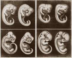
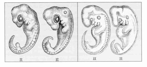
Page 48: "In the first edition of this book, Haeckel used the same drawing, only he reproduced it three times claiming that it represented the embryos of three different creatures. The famous naturalist Francis Agassiz spotted this farce, and made critical comments in the margin of his personal copy of this book."
NOTE: The article contains a minor error on page 48, where it is stated that the three animals were labeled as a dog, chicken, and turtle, whereas beneath the picture on page 46, it is stated that that these were a dog, pig, and turtle.
[1549] Book: The History of Creation: Or The Development of the Earth and Its Inhabitants by the Action of Natural Causes. By Ernst Haeckel. Translated by E. Ray Lankester. Volume 1. D. Appleton and Company, 1879. From the fourth German edition of the book entitled Naturliche Schöpfungsgeschichte, 1873. The first edition was published in 1868.
Page 294: "The facts of embryology alone would be sufficient to solve the question of man's position in nature.... Look attentively at and compare the eight figures which are represented on the adjoining Plates II. and III., and it will be seen that the philosophical importance of embryology cannot be too highly estimated."
NOTE: The figures referenced above appear on the unnumbered pages following page 306:
Page 307: "Everyone surely knows the gill-arches of fish, those arched bones that lie behind one another... and which support the gills, the respiratory organs of the fish. ... Now these gills arches originally exist exactly the same in man (D), in dogs (C), in fowls (B), and in tortoises (A), as well as in all other vertebrate animals."
[1550] Book: The History of Creation: Or The Development of the Earth and Its Inhabitants by the Action of Natural Causes. By Ernst Haeckel. Translated by E. Ray Lankester. Volume 1. D. Appleton and Company, 1879. From the fourth German edition of the book entitled Naturliche Schöpfungsgeschichte, 1873. The first edition was published in 1868. Pages 308-9.
Immediately preceding the cited quote are these words:
Most persons even now refuse to acknowledge the most important deduction of the theory of Descent, that is, the palaeontological development of man from ape-like, and through them from still lower, mammals, and consider such a transformation of organic form as impossible. But, I ask, are the phenomena of the individual development of man, the fundamental features of which I have here given, in any way less wonderful?
[1551] Book: The Descent of Man, And Selection in Relation to Sex. By Charles Darwin. Second edition. John Murray, 1874. 1890 reprint. First published in 1871. Pages 24-25:
With respect to development, we can clearly understand, on the principle of variations supervening at a rather late embryonic period, and being inherited at a corresponding period, how it is that the embryos of wonderfully different forms should still retain, more or less perfectly, the structure of their common progenitor. No other explanation has ever been given of the marvelous fact that the embryos of a man, dog, seal, bat, reptile, etc, can at first hardly be distinguished from each other.
[1552] Book: The History of Creation: Or The Development of the Earth and Its Inhabitants by the Action of Natural Causes. By Ernst Haeckel. Translated by E. Ray Lankester. Volume 1. D. Appleton and Company, 1879. From the fourth German edition of the book entitled Naturliche Schöpfungsgeschichte, 1873. The first edition was published in 1868. Pages 309-310:
Verily, if we compare those two series of development with one another, and ask ourselves which of the two is the more wonderful, it must be confessed that ontogeny, or the short and quick history of the development of the individual, is much more mysterious than phylogeny, or the long and slow history of development of the tribe. For one and the same grand change of form is accomplished by the latter in the course of many thousands of years, and by the former in the course of a few months. Evidently this most rapid and astonishing transformation of the individual in ontogenesis, which we can actually point out at any moment by direct observation, is in itself much more wonderful and astonishing than the corresponding, but much slower and gradual transformation which the long chain of ancestors of the same individual has gone through in phylogenesis.
I have endeavored in the second volume of the "General Morphology, to establish this theory in detail, as I consider it exceedingly important. As I have there shown, ontogenesis, or the development of the individual, is a short and quick repetition (recapitulation) of phylogenesis, or the development of the tribe to which it belongs, determined by the laws of inheritance and adaptation; by tribe I mean the ancestors which form the chain of progenitors of the individual concerned. ... In this intimate connection of ontogeny and phylogeny, I see one of the most important and irrefutable proofs of the Theory of Descent. No one can explain these phenomena unless he has recourse to the laws of Inheritance and Adaptation; by these alone are they explicable.
[1553] Textbook: Developmental Biology. By Werner A. Müller. English translation. Springer-Verlag, 1997.
Page 124: "Ernst Haeckel ... drafted his much-disputed "biogenetic law." Actually, this "law" is a hypothesis.... In its succinct and catchy form, the law states that "ontogeny recapitulates phylogeny" in a condensed and abbreviated way."
[1554] Book: Fundamentals of Comparative Embryology of the Vertebrates. By Alfred F. Huettner. Revised edition. Macmillan Company, 1949. First edition published in 1941.
Page 6 states that the recapitulation theory is "usually ascribed to Häckel."
Page 39 states the recapitulation theory was formulated by Fritz Muller in 1863 and forecast by von Baer in 1828.
NOTE: See the next three sources, which show that a comparable theory was articulated and popularized by Chambers before Müller, and its roots can be traced back to at least 1811 in a writing of Meckel.
[1555] Paper: "The discovery of the mammalian egg and the foundation of modern embryology." By George Sarton. Isis, November 1931. Pages 315-330.
Page 326: "[T]he theory that the embryonic development of each creature is a brief recapitulation of its ancestral history. That theory was elaborated by FRITZ MÜLLER (1821-97) in his book Für Darwin (Leipzig, 1864), and popularized by HAECKEL. (9)"
Note (9) states that the author has traced the idea at least as far back as 1811 in a writing of Johann Friedrich Meckel. Also, he notes that the general concept "perhaps" appears in a writing published in 1793, and he states: "I wonder if that had much influence in the development of the doctrine...."
[1556] Book: Vestiges of the Natural History of Creation. By Anonymous [Robert Chambers]. John Churchill, 1844. Electronic edition prepared by Robert Robbins. http://www.esp.org/books/chambers/vestiges/facsimile/
Pages 198-9:
We have yet to advert to the most interesting class of facts connected with the laws of organic development. It is only in recent times that physiologists have observed that each animal passes, in the course of its germinal history, through a series of changes resembling the permanent forms of the various orders of animals inferior to it in the scale. ... Nor is man himself exempt from this law. His first form is that which is permanent in the animalcule. His organization gradually passes through conditions generally resembling a fish, a reptile, a bird, and the lower mammalia, before it attains its specific maturity.
Page 212:
It has been seen that, in the reproduction of the higher animals, the new being passes through stages in which it is successively fish-like and reptile-like. But the resemblance is not to the adult fish or the adult reptile, but to the fish and reptile at a certain point in their fetal progress; this holds true with regard to the vascular, nervous, and other systems alike.
[1557] Book: Organic Evolution as the Result of the Inheritance of Acquired Characters According to the Laws of Organic Growth. By G. H. Theodor Eimer (Professor of Zoology and Comparative Anatomy in Tübingen). Translated by J. T. Cunningham. Macmillan and Co., 1890. Page 30:
The highest animals briefly repeat in their ontogeny the whole series of their ancestors (biogenetic law) as stages of growth. ... Thus the facts established by me afford at the same time provide a new and complete confirmation of the biogenetic law. Varieties and species are therefore in reality nothing but groups of forms standing at different stages of evolution....
[1558] Book: The Shape of Life: Genes, Development, and the Evolution of Animal Form. By Rudolf A. Raff. University of Chicago Press, 1996.
Page 2: "Darwin's most forceful adherent was the German zoologist Ernst Haeckel.... In 1866, he propounded the famous and overwhelmingly influential biogenetic law, which states that ontogeny (the development of the individual) results from phylogeny (the evolutionary history of the lineage)."
NOTE: This author and the one in the next note do not accept this supposed "law," but explain that it was very popular and commonly referred to as a "law."
[1559] Book: Comparative Embryology of the Vertebrates. By Olin E. Nelsen (Department of Zoology, University of Pennsylvania). Blakiston Company, 1953.
Page 348: "Many have been the supporters of the biogenetic law, and for a long time it was one of the most popular theories of biology."
[1560] Article: "Life After Death Declared Proved By Evolution." By George MacAdam. New York Times, December 14, 1913.
But, now, here is a man, Dr. J. Leon Williams, Fellow of the Anthropological Institute of Great Britain and Ireland, who has spent his life studying the family tree of prehistoric man....
The evidence of man's ascent to be found in his prehistoric remains, and in almost every part of his own body, is so overwhelming as to be almost beyond discussion. ...
Take just a few of the evidences that exist in his own body. ...
It is perfectly well known that the human embryo in its development passes through the entire evolutionary process of the vertebrates.
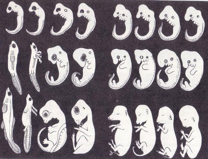
[1561] Book: The Evolution of Man: A Popular Exposition of the Principal Points of Human Ontogeny and Phylogeny. By Ernst Haeckel. Volume 1. D. Appleton and Company, 1896. Translated from the German book entitled Anthropogenie, which was first published in 1874. Page xix [the first page of the preface to the first edition]:
Few educated men have any suspicion of the fact, that these human embryos conceal a greater wealth of important truths, and form a more abundant source of knowledge than is afforded by the whole mass of most other sciences and of all so-called "revelations."
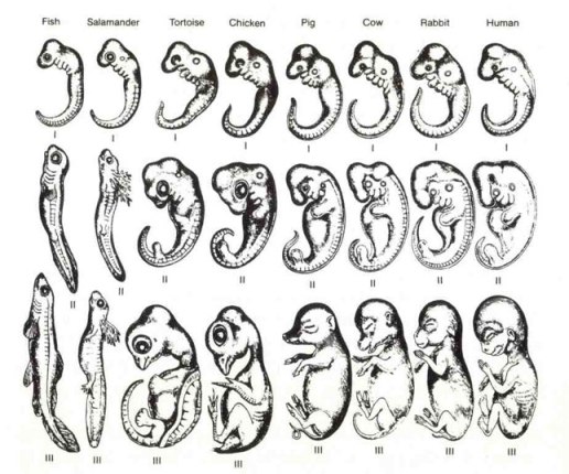
Pages 360-1:
A careful study and thoughtful comparison of the embryos of Man and other Vertebrates in this stage of development is very instructive, and reveals to the thoughtful many profounder mysteries and weightier truths than are to be found in the so-called "revelations" of all the ecclesiastical religions of the world. Compare, for instance, carefully and attentively the three consecutive stages of development ... [of the Fish, Salamander, Tortoise, Chick, Hog, Calf, Rabbit and Man]. In the first stage (upper Row of Section I.), in which the head with the five brain-bladders, and the gill-arches are indeed begun, though the limbs are still entirely wanting, the embryos of all Vertebrates from Fish to Man differ not at all, or only in non-essential points. ... The significance of such facts as these cannot be over-estimated. ...
... For the wonderful and comprehensive harmony between the individual evolution of Man and that of other Vertebrates is only explicable by assuming the descent of these from a common parent-form.
[1562] Book: The Evolution of Man: A Popular Exposition of the Principal Points of Human Ontogeny and Phylogeny. By Ernst Haeckel. Volume 1. D. Appleton and Company, 1896. Translated from the German book entitled Anthropogenie, which was first published in 1874.
Drawing appears on the unnumbered pages after page 362.
[1563] Article: "Sedgwick, Adam." Encyclopedia Britannica Ultimate Reference Suite 2004.
NOTE: The Adam Sedgwick who wrote this paper should not be confused with his great-uncle of the same name, who was a creationist (see citation 1661). As this article explains, both Sedgwicks were accomplished scientists.
[1564] Paper: "On the law of development commonly known as von Baer's law; and on the significance of ancestral rudiments in embryonic development." By Adam Sedgwick. Quarterly Journal of Microscopical Science, April 1, 1894. Pages 35-52. http://jcs.biologists.org/cgi/reprint/s2-36/141/35
Page 36: "The examples I have chosen are the fowl and dog-fish. ... There is no stage of development in which the unaided eye would fail to distinguish between them with ease.... A blind man could distinguish between them.
[1565] Paper: "On the law of development commonly known as von Baer's law; and on the significance of ancestral rudiments in embryonic development." By Adam Sedgwick. Quarterly Journal of Microscopical Science, April 1, 1894. Pages 35-52. http://jcs.biologists.org/cgi/reprint/s2-36/141/35
Footnote 1:
I do not feel called upon to characterize the accuracy of the drawings of embryos of different classes of the Vertebrata given by Haeckel in his popular works, and reproduced by Romanes and, for all that I know, other popular exponents of the evolution theory. As a sample of their accuracy, I may refer the reader to the varied position of the auditory sac in the drawings of the younger embryos.
[1566] Paper: "On the law of development commonly known as von Baer's law; and on the significance of ancestral rudiments in embryonic development." By Adam Sedgwick. Quarterly Journal of Microscopical Science, April 1, 1894. Pages 35-52. http://jcs.biologists.org/cgi/reprint/s2-36/141/35
Pages 38-39:
If v. Baer's law has any meaning at all, surely it must imply that animals so closely allied as the fowl and duck would be indistinguishable in the early stages of development ... yet I can distinguish a fowl and a duck embryo on the second day by the inspection of a single transverse section through the trunk.... But it is not necessary to emphasize further these embryonic differences; every embryologist knows that they exist and could bring forward innumerable instances of them. I need only say with regard to them that a species is distinct and distinguishable from its allies from the very earliest stages all through the development, although these embryonic differences do not necessarily implicate the same organs as do the adult differences.
[1567] Paper: "On the law of development commonly known as von Baer's law; and on the significance of ancestral rudiments in embryonic development." By Adam Sedgwick. Quarterly Journal of Microscopical Science, April 1, 1894. Pages 35-52. http://jcs.biologists.org/cgi/reprint/s2-36/141/35
Pages 41-2:
In fact the balance of evidence appears to me to point most clearly to the fact that the tendency in embryonic development is to directness and abbreviation and to the omission of ancestral stages of structure, and that variations do not merely affect the not-early period of life where they are of immediate functional importance to the animal, but, oh the contrary, that they are inherent in the germ and affect more or less profoundly the whole of development. I am well aware that in holding this opinion I am running counter to the great authority of Darwin.
[1568] Obituary Notice: "Erik Nordenskiöld (1872-1933)." By Nils V. Hofsten. Pages 103-6. Isis, November 1947.
Page 103 notes that his uncle was a famous arctic explorer of the same name.
Page 105: "A German edition followed in 1926, a Finnish edition in 1927-1929, and an English edition in 1929. ... [I]t is said that he spent most of his time in the library of the University studying old authors and arrived often at the lectures with a heavy load of bulky folios from which he recited appropriate fragments...."
On page 105, Hofsten states that the "merits and the positive influence" of Haeckel "seem to be a little undervalued" in this work. {Based on what we know today, Nordenskiöld was charitable in his treatment of Haeckel (see citation 1572).}
[1569] Home page: "Department of Zoology at Michigan State University." Accessed September 9, 2007 at http://www.zoology.msu.edu/
"Zoology is the branch of natural science that deals with the integrative study of animal biology."
[1570] Web Page: "History of Zoology at Southern -- The Period From 1915-1940." Department of Zoology, College of Science at Southern Illinois University. Last updated February 14, 2008. Accessed December 12, 2009 at http://www.science.siu.edu/zoology/history/1915-1940.htm
"A course on the history of biology used what was then a relatively new book on the subject by Erik Nordenskiold. No scholar has been able to write a comparable history in the past 60 years and thus the History of Biology remains the text of choice for courses on the background of our discipline."
[1571] See pages 212-213 of Rational Conclusions and citations 1589-1599, 1624-1631.
[1572] Book: The History of Biology: A Survey. By Erik Nordenskiöld. Tudor Publishing, 1946. Translated from the Swedish volume entitled Biologins Historia, 1920-24. Page 517:
Being designed exclusively to prove one single assertion, his illustrations were naturally extremely schematic and without a trace of scientific value, sometimes indeed so far divergent from the actual facts as to cause him to be accused of deliberate falsification – an accusation that a knowledge of his character would have at once refuted.1 ...
1 It is nevertheless difficult to understand such an action as this: allowing in his Natürliche Schöpfungsgeschichte (ed. i, p. 242) the same cliché, reproduced three times, to represent the egg of a man, an ape, and a dog. This absurdity was removed from subsequent editions, albeit only after Haeckel had rewarded with abuse those who pointed out the fact; and the incident was ever afterwards a theme on which his enemies constantly harped.
Page 522: "[E]verything of value in his utterances has become permanent, while his blunders have been forgotten, as they deserve."
NOTE: Nordenskiöld criticizes Darwin and his views of heredity on pages 469, 477-8, 573, and 616, but does not contest evolution and implies acceptance of it with this statement on page 573: "[F]ormerly one sought in the phenomena of life manifestations of a divine creator; when this was no longer perceivable, one had to look for a material creative power—it was difficult to realize that evolution is a part of life itself."
[1573] See citation 1548 for another example of Haeckel reproducing the exact same drawing and claiming it represents the embryos of different creatures.
[1574] Book: Comparative Embryology of the Vertebrates. By Olin E. Nelsen (Department of Zoology, University of Pennsylvania). Blakiston Company, 1953.
Page 530: "A, D, H, M, and Q show primitive embryonic body form in the developing shark, rock fish, frog, chick, and human." [The sketch is on page 531.]
[1575] Textbook: Embryology: Constructing the Organism. Edited by Scott F. Gilbert & Anne M. Raunio. Sinauer Associates, 1997.
Page x states the book is intended for college sophomores. Alongside a drawing derived from Haeckel's, page 384 states that "at an early stage all vertebrate embryos are very similar and exhibit the general features of the vertebrate subphylum (I) ... (From Romanes 1901.)" The top row of the drawing is labeled "(I)". That Romanes's drawing is derived from Haeckel's is shown by the citation below.
[1576] Paper: "On the law of development commonly known as von Baer's law; and on the significance of ancestral rudiments in embryonic development." By Adam Sedgwick. Quarterly Journal of Microscopical Science, April 1, 1894. Pages 35-52. http://jcs.biologists.org/cgi/reprint/s2-36/141/35
Page 36: "I do not feel called upon to characterize the accuracy of the drawings of embryos of different classes of the Vertebrata given by Haeckel in his popular works, and reproduced by Romanes and, for all that I know, other popular exponents of the evolution theory."
[1577] Paper: "There is no highly conserved embryonic stage in the vertebrates: implications for current theories of evolution and development." By Michael K. Richardson and others. Journal of Anatomy and Embryology, July, 1997. Pages 91-106. http://www.springerlink.com/content/1cf2gngc2qee6efp/...
Page 91:
Some authors have suggested that members of most or all vertebrate clades pass through a virtually identical, conserved stage. This idea was promoted by Haeckel, and has recently been revived in the context of claims regarding the universality of developmental mechanisms. ... In view of the current widespread interest in evolutionary developmental biology, and especially in the conservation of developmental mechanisms, re-examination of the extent of variation in vertebrate embryos is long overdue.
Page 92: "One puzzling feature of the debate in this field is that while many authors have written of a conserved embryonic stage, no one has cited any comparative data in support of the idea. It is almost as though the phylotypic stage is regarded as a biological concept for which no proof is needed."
Page 93: "The idea of a phylogenetically conserved stage has regained popularity in recent years."*
Page 94 lists 39 different species used in the study.
Page 95: "Haeckel's drawings of embryos at tailbud stages are widely used in support of this hypothesis."
NOTE: For an example, see the citation below.
[1578] Textbook: Embryology: Constructing the Organism. Edited by Scott F. Gilbert & Anne M. Raunio. Sinauer Associates, 1997. Page 384:

NOTE: Adjacent to this drawing, the book states that "at an early stage all vertebrate embryos are very similar.... This has since been termed the phylotypic stage."
[1579] Book: Endless Forms Most Beautiful. By Sean B. Carroll. W. W. Norton & Company, 2005.
Page 9: "The comparison of developmental genes between species became a new discipline at the interface of embryology and evolutionary biology—evolutionary developmental biology, or 'Evo-Devo' for short."
[1580] Paper: "Inverting the hourglass: quantitative evidence against the phylotypic stage in vertebrate development." By Olaf R. P. Bininda-Emonds & others. Proceedings of the Royal Society: Biological Sciences, January 20, 2003. http://www.pubmedcentral.nih.gov/articlerender.fcgi?artid=1691251
Page 341:
The concept of a phylotypic stage, when all vertebrate embryos show low phenotypic diversity, is an important cornerstone underlying modern developmental biology. Many theories involving patterns of development, developmental modules, mechanisms of development including developmental integration, and the action of natural selection on embryological stages have been proposed with reference to the phylotypic stage.
[1581] Paper: "There is no highly conserved embryonic stage in the vertebrates: implications for current theories of evolution and development." By Michael K. Richardson and others. Journal of Anatomy and Embryology, July, 1997. Pages 91-106. http://www.springerlink.com/content/...
[1582] On September 28, 2009, I wrote to Springer Publishing (which publishes the Journal of Anatomy and Embryology), requesting permission to use the embryo photos. On October 27th, I received a reply stating that Springer does not own the copyright to these photos and suggesting I contact the Hubrecht Laboratory or authors of the paper. Given that Michael Richardson is the lead author of the paper, has been involved with the Hubrecht Laboratory (http://www.mk-richardson.com/index.php?...), and previously gave permission to use some of these photos to Creation Ministries International (http://creation.com/fraud-rediscovered), I wrote to him on October 28, 2009 requesting permission to use the photos. I have not yet received a reply.
[1583] Article: "An embryonic liar." By Nigel Hawkes. London Times, August 11, 1997. Page 14:
Dr
Michael Richardson, has shown that even
this, Haeckel's last bequest to science, is
deeply flawed.
"This is one of the worst cases of
scientific fraud. It’s shocking to find that
somebody one thought was a great scientist
was deliberately misleading. It makes me
angry." ...
... "What he did was to take a human embryo
and copy it, pretending that the salamander
and the pig and all the others looked the
same at the same stage of development. There
is only one word for this, and Dr Richardson
doesn't flinch from using it. "These are
fakes. In the paper, we call them
'misleading and inaccurate', but that is
just polite scientific language."
[1584] Paper: "There is no highly conserved embryonic stage in the vertebrates: implications for current theories of evolution and development." By Michael K. Richardson and others. Journal of Anatomy and Embryology, July, 1997. Pages 91-106. http://www.springerlink.com/content/...
Page 98: "The zebrafish at 0.9 mm was the smallest embryo included in this review."
Page 103: "Size is another parameter which varies tremendously between tailbud embryos – from 700 μm [micrometers - one millionth of a meter] in the scorpion fish [not reviewed in this study] to 9.25 mm in the mudpuppy."
Page 98: "Our series varies in size from the small embryo of the striped chorus frog (Pseudacris triseriata) at 1.5 mm, to the large embryo of the mudpuppy (Necturus maculosus), which has a greatest length of 9.25 mm."
[1585] Article: "An embryonic liar." By Nigel Hawkes. London Times, August 11, 1997. Page 14.
[1586] See pages 201-203 & 212-214 of Rational Conclusions.
[1587] Search at http://alacarte.lexisnexis.com on August 15, 2007. Three separate searches were performed with the terms (1) Richardson AND Haeckel, (2) Haeckel AND embryos, and (3) "Ernst Haeckel." The date range was from July 1, 1997 – July 1, 1998. (The study was published in July 1997). I examined the summary of each result to see if there was reference to the study or the drawings. Although unlikely, there might be mention of this topic buried in other stories, but this clearly does not constitute a story about the topic.
[1588] Search at http://alacarte.lexisnexis.com on August 15, 2007. The search was performed for the word Tiktaalik. The date range was from April 1, 2006 – April 1, 2007. (The study was published on April 6, 2007 in the journal Nature). I examined the summary of each result to see if there was reference to the study or the fossil. Thus, there might be mention of this topic buried in other stories, but this clearly does not constitute a story about the topic. In addition to the publications mentioned in the main text, there were also articles appearing in these publications:
| The Advertiser (Australia) | The Age (Melbourne, Australia) |
| Akron Beacon Journal (Ohio) | The Australian |
| Australian Broadcasting Corporation (ABC) | Bangor Daily News (Maine) |
| Belfast Telegraph | Belleville News-Democrat (Illinois) |
| Birmingham Post | Bismarck Tribune |
| Bradenton Herald (Florida) | Brantford Expositor (Ontario) |
| Broadcast News (Canada) | Buffalo News (New York) |
| Calgary Herald (Alberta) | Canadian Press (CP) |
| Charlotte Observer (North Carolina) | Chinadaily.com.cn |
| Christian Science Monitor | Cincinnati Enquirer (Ohio) |
| Cincinnati Post (Ohio) | Columbus Dispatch (Ohio) |
| Commercial Appeal (Memphis, TN) | Copley News Service |
| Daily Herald-Tribune (Grande Prairie, Alberta) | Daily Mail (London) |
| Daily Telegraph (Australia) | Daily Telegraph (London) |
| Daily Yomiuri (Tokyo) | Denver Post |
| Economist | Edmonton Journal (Alberta) |
| Edmonton Sun (Alberta) | Evening Standard (London) |
| EWorldWire | The Express |
| Facts on File World News Digest | Financial Times (London, England) |
| The Gazette (Montreal) | Globe and Mail (Canada) |
| Grand Rapid Press (Michigan) | Guardian (London) |
| Guardian Weekly | Hamilton Spectator (Ontario, Canada) |
| Hindustan Times | Houston Chronicle |
| The Independent (London) | International Herald Tribune |
| Irish Independent | Irish Times |
| Kamloops Daily News (British Columbia) | Kansas City Star |
| Kingston Whig-Standard (Ontario) | Knight Ridder Washington Bureau |
| Lexington Herald Leader (Kentucky) | London Free Press (Ontario) |
| Miami Herald | Monterey County Herald (California) |
| Myrtle Beach Sun-News (South Carolina) | Nanaimo Daily News (British Columbia) |
| National Post (f/k/a The Financial Post) (Canada) | Natural History |
| Niagara Falls Review (Ontario) | Ottawa Citizen |
| Press Association Newsfile | Prince George Citizen (British Columbia) |
| Prince Rupert Daily News (British Columbia) | The Record (Kitchener-Waterloo, Ontario) |
| Religion News Service | Richmond Times Dispatch (Virginia) |
| Roanoke Times (Virginia) | Rocky Mountain News (Denver, CO) |
| Sarasota Herald-Tribune (Florida) | Sault Star (Sault Saint Marie Ontario) |
| Scripps Howard News Service | Simcoe Reformer (Ontario, Canada) |
| St. John’s Telegram (Newfoundland) | St. Louis Post-Dispatch (Missouri) |
| St. Petersburg Times (Florida) | Star Phoenix (Saskatoon, Saskatchewan) |
| The State (Columbia, South Carolina) | States News Service |
| Statesman (India) | Sydney Morning Herald (Australia) |
| Sydney MX (Australia) | Times Colonist (Victoria, British Columbia) |
| Toronto Star | Toronto Star |
| US Fed News | Vancouver Province (British Columbia) |
| Vancouver Sun (British Columbia) | Voice of America News |
| The West Australian (Perth) | Wichita Eagle (Kansas) |
| Windsor Star (Ontario) | York Dispatch (Pennsylvania) |
[1589] Book: Elements of Molecular Neurobiology. By C. U. M. Smith. Third edition. John Wiley & Sons, 2002.
Page 419: "the early embryos of a wide variety of vertebrates look very alike."
Page 420 shows this drawing:

[1590] Textbook: Before We Are Born: Essentials of Embryology and Birth Defects. By Keith L. Moore & T.V.N. Persaud. Saunders, 2003. Sixth edition.
Page 152: "At about four weeks of development, the head and neck regions of the human embryo somewhat resemble those regions of a fish embryo at a comparable stage of development."
[1591] Textbook: Langman's Medical Embryology. By T. W. Sadler. Ninth edition. Lippincott Williams & Wilkins, 2004.
The rear cover states this book is "Recognized as the classic textbook in embryology...."
Page 364: "Although development of pharyngeal arches, clefts and pouches resembles formation of gills in fishes and amphibia, in the human embryo real gills (branchia) are never formed."
[1592] Textbook: Evolution. By Monroe W. Strickberger. Third edition. Jones and Bartlett, 2000.
Page 44 shows a drawing derived from Haeckel's and states: "Note that each of the embryos begins with a similar number of pharyngeal (gill) arches (pouches below the head) and a similar vertebral column."
[1593] Article: "Fetus." Black's Medical Dictionary. Edited by Gordon Macpherson. 39th edition. Madison Books, 1999. Pages 202-3. Page 203:
From two weeks after conception onward, the various organs and limbs appear and grow, the name of embryo being applied to the developing being while almost indistinguishable in appearance from the embryo of other animals, till the middle of the second month, when it begins to show a distinctly human form. After this stage it is called the fetus.
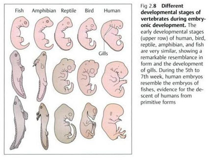
[1594] Book: What Evolution Is. By Ernst Mayr. Basic Books, 2001.
Page 27: "An early human embryo, for instance is very similar not only to the embryos of other mammals (dog, cow, mouse), but in its early stages even to those of reptiles amphibians and fishes (Fig 2.8).
Page 28, Figure 2.8:
[1595] Article: "Inside The Womb." By J. Madeleine Nash. Time, November 11, 2002. Pages 68-78.
Page 71: "40 days - At this point, a human embryo looks no different from that of a pig, chick or elephant. All have a tail, a yolk sac and rudimentary gills."
[1596] Article: Was Darwin Wrong? By David Quammen. National Geographic, November 2004 (cover story). http://ngm.nationalgeographic.com/ngm/0411/feature1/fulltext.html
"Embryology too involved patterns that couldn't be explained by coincidence. Why does the embryo of a mammal pass through stages resembling stages of the embryo of a reptile?"
[1597] Book: The Tapir's Morning Bath: Mysteries of the Tropical Rain Forest and the Scientists Who Are Trying to Solve Them. By Elizabeth Royte. Houghton Mifflin, 2001.
Pages 182-3: "In humans, Haeckel's law is seen in action as embryos pass through stages reminiscent of fish, amphibians, and reptiles."
[1598] Book: Growth and Development. By Virginia B. Silverstein, Alvin Silverstein & Laura Silverstein Nunn. Twenty-First Century Books, 2008.
Page 69: "All vertebrates look like one another in certain stages of their early development. A mammal goes through fishlike and reptilelike stages during its development before birth."
[1599] Web Page: "Meet the Author: "Alvin, Robert, and Virginia Silverstein." Houghton Mifflin Company. Accessed September 15, 2007 at http://www.eduplace.com/kids/hmr/mtai/silverstein.html
"Alvin Silverstein and Virginia Silverstein ... have published more than 160 books on science and health topics."
[1600] Letter to the editor: "Haeckel, Embryos and Evolution." By Michael K. Richardson and others. Science, May 15, 1998. Pages 983 ff.
[1601] Article: "An embryonic liar." By Nigel Hawkes. London Times, August 11, 1997. Page 14:
Dr
Michael Richardson, has shown that even
this, Haeckel's last bequest to science, is
deeply flawed.
"This is one of the worst cases of
scientific fraud. It’s shocking to find that
somebody one thought was a great scientist
was deliberately misleading. It makes me
angry." ...
... "What he did was to take a human embryo
and copy it, pretending that the salamander
and the pig and all the others looked the
same at the same stage of development. There
is only one word for this, and Dr Richardson
doesn't flinch from using it. "These are
fakes. In the paper, we call them
'misleading and inaccurate', but that is
just polite scientific language."
[1602] Book: In Search of Deep Time: Beyond the Fossil Record to a New History of Life. The Free Press, 1999.
Page 75: "In vertebrates, the notochord forms a kind of scaffolding for the bony vertebral discs, which replace and supplant it."
[1603] Entry: "notochord." American Heritage Dictionary of Science. Edited by Robert K. Barnhart. Houghton Mifflin, 1986.
Page 440: "In the higher chordates the notochord is present in the embryo only since it is replaced by the bony vertebral column in the adult form (Winchester, Zoology)."
[1604] Textbook: Langman's Medical Embryology. By T. W. Sadler. Ninth edition. Lippincott Williams & Wilkins, 2004.
Page 364: "Pharyngeal arches not only contribute to the formation of the neck, but also play an important part in formation of the face."
[1605] Paper: "There is no highly conserved embryonic stage in the vertebrates: implications for current theories of evolution and development." By Michael K. Richardson and others. Journal of Anatomy and Embryology, July, 1997. Pages 91-106. http://www.springerlink.com/content/...
Page 91: "We find that embryos at the tailbud stage – thought to correspond to a conserved stage – show variations in form due to allometry, heterochrony, and differences in body plan and somite number. These variations foreshadow important differences in adult body form."
[1606] Paper: "Inverting the hourglass: quantitative evidence against the phylotypic stage in vertebrate development." By Olaf R. P. Bininda-Emonds & others. Proceedings of the Royal Society: Biological Sciences, January 20, 2003. http://www.pubmedcentral.nih.gov/...
Page 344:
Support for the hourglass definition of the phylotypic stage derives largely from subjective statements about the overall similarity of embryos of different species, usually based on an examination of pictures of embryos and not from rigorous character-based data analysis. {Note from this context that this is a reference to the tailbud stage.} ...
... Shared features undoubtedly exist during the mid-embryonic period, but those used to support the phylotypic stage are often defined so coarsely as to obscure potential variation between species. For instance, the statement that vertebrate embryos all possess a heart during the phylotypic [tailbud] stage (Kimmel et al. 1995) ignores important variation in how the heart is formed (see Richardson 1995) as well as the existence of heterochrony, which can result Proc. R. Soc. Lond. B (2003) in the heart being in different stages of its development when other key characters are all present (which may themselves be at varying stages in their development).
[1607] Article: "Abscheulich! (Atrocious!)" By Stephen J. Gould. Natural History, March 2000. Pages 42-49. Page 48.
[1608] Same as above.
[1609] Book: Fundamentals of Comparative Embryology of the Vertebrates. By Alfred F. Huettner. Revised edition. Macmillan Company, 1949. First edition published in 1941.
Page 39: "As a "law," this principle has been questioned. It has been subjected to careful scrutiny and has been found wanting. There are too many exceptions to it. However, there is no doubt that it contains some truth and that it is of value to the student of embryology."
[1610] Book: Old Fourlegs: The Story of the Coelacanth. By J. L. B. Smith. Longman's, Green and Co, 1956.
Page 236: "The development of embryos is a most fascinating study, for it has been observed that many show characters of the earliest forms of life from which the creatures have evolved."
Page 246: "Most people know that a developing embryo shows features which are believed to be clues to ancestral forms."
[1611] Book: The Evolutionary Process: A Critical Review of Evolutionary Theory. By Verne Grant (Ph.D. in botany and genetics from Berkeley, Professor of Botany at The University of Texas at Austin). Columbia University Press, 1985.
Page 364: "Therefore Haeckel's conclusion is not a universal law, nor is it discredited, but it stands as a useful generalization."
[1612] Book: Ontogeny and Systematics. Edited by C.J. Humphries. Columbia University Press, 1988. Chapter 7: "Epigenetics." By S. Løvtrup (former Professor of the Department of Zoophysiology, University of Umeå, Sweden).
Page 191: "This recapitulation is not an adult recapitulation as implied by the biogenetic law, nor is really a true embryonic recapitulation, even if it is closer to the latter than to the former."
Pages 194-5: "According to the traditional recapitulation theory this is most unfortunate, but if, as suggested here, recapitulation begins only during gastrulation, such differences are less important."
Page 196:
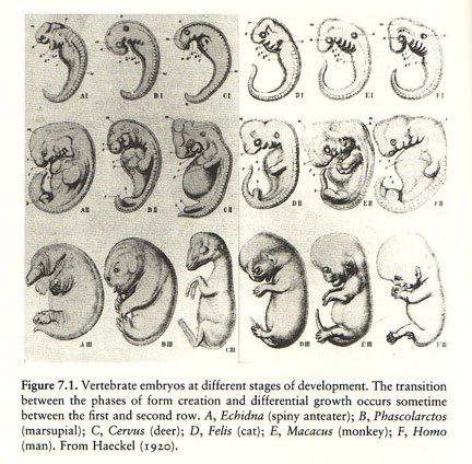
NOTE: Page 225 lists the source of this drawing as the 12th edition of Haeckel's Natürliche Schöpfungsgeschichte, 1920. Haeckel died in 1919. This drawing contains some alleged embryos not included in Haeckel's earlier drawings. Take special notice of the cat, a sketch of which is shown on page 211 of Rational Conclusions.
Page 224: "If this assertion is accepted then ontogenetic development will constitute a recapitulation of the course of phylogenetic evolution."
[1613] Book: Genetics, Paleontology, and Macroevolution. By Jeffrey Levinton (State University of New York at Stony Brook). Cambridge University Press, 1988. Page 266:
The universality of the biogenetic law was refuted by the demonstration of rearrangements of the order of appearance of structures between ancestor and descendant.... The law still strongly influences our thinking however. ... [D]iscussions above tend to suggest that ontogeny and phylogeny might very well be intimately related, perhaps sometimes to the degree that the biogenetic law may hold.
[1614] Textbook: Biology. By Peter H. Raven & George B. Johnson. Fifth edition (customized). McGraw-Hill, 1999.
Page 416: "Some of the strongest anatomical evidence supporting evolution comes from comparisons of how organisms develop. In many cases, the evolutionary history of an organism can be seen to unfold during its development, with the embryo exhibiting characteristics of the embryos of its ancestors (figure 20,18)."
[1615] Book: What Evolution Is. By Ernst Mayr. Basic Books, 2001. Pages 29-30:
Ontogeny is the recapitulation of phylogeny" obviously went too far, because at no stage of its development does a mammalian embryo look like an adult fish. Yet, in certain instances as in the gill pouches {claims about gill pouches in mammals are refuted on pages 215-216 of Rational Conclusions}, the mammalian embryo does indeed recapitulate the ancestral condition. And such cases of recapitalization are by no means rare. ... [A]ll terrestrial vertebrates (tetrapods) develop gill arches at a certain stage in their ontogeny.
[1616] Book Review: "How to Build a Dinosaur by Jack Horner and James Gorman." By Jeff Hecht. New Scientist, February 25, 2009. http://www.newscientist.com/article/...
He [Jack Horner] wants to alter the embryological development of chickens, which are living descendants of dinosaurs. His idea comes from the fertile field of "evo-devo", which focuses on how evolution affects the way animals develop from fertilized eggs. Look closely at a developing embryo and you can see some ancestral forms briefly appear. Birds, for example, start to develop tails, then convert the would-be-tail into a pygostyle, a bony lump at the base of the spine which holds the tail feathers.
[1617] Book: Evolution and Genetics. By David J. Merrell. Holt, Rinehart and Winston, 1962.
Page 88: "There is a germ a truth in the biogenetic law even though it is demonstrably false if taken too literally...."
Page 93: "The recapitulation theory of Haeckel, as originally stated, represents an oversimplification of the facts, for the developing embryo does not recapitulate the adult stages of its ancestors. Rather, the embryo will in most instances show more resemblance to the embryos of ancestral or related groups than it will to their adult forms."
[1618] Textbook: Developmental Biology. By Werner A. Müller. English translation. Springer-Verlag, 1997. Pages 124-5:
Haeckel's biogenetic law merits acknowledgement as it points to the evolutionary context of developmental biology, but it must be corrected: each organism's ontogeny does not repeat phylogeny of a species but rather previous ontogenies. In each generation all species recapitulate their own ontogeny, which, compared with the ontogeny of related species, is more or less modified. On the other hand, all vertebrates pass through a highly conserved common stage that displays a uniform basic body architecture characteristic of all vertebrates. Therefore, the biogenetic law is valid if it is modified by stating that all vertebrates recapitulate certain embryonic states of their ancestors—in particular, a common phylotypic stage.
[1619] Textbook: Asking About Life. By Allan J. Tobin & Jennie Dusheck. Third Edition. Brooks Cole, 2004.
Page 317: "The embryos of developing organisms frequently pass through stages that resemble the embryos of organisms from which they evolved, a fact consistent with the theory of evolution."
[1620] Paper: "Punctuated Equilibria: The Tempo and Mode of Evolution Reconsidered." By Stephen Jay Gould and Niles Eldredge. Paleobiology, Spring 1977. Pages 115-151. Page 147:
At the higher level of evolutionary transition between basic morphological designs, gradualism has always been in trouble, though it remains the "official" position of most Western evolutionists. Smooth intermediates between Baupläne [body plans] are almost impossible to construct, even in thought experiments; there is certainly no evidence for them in the fossil record (curious mosaics like Archaeopteryx do not count). ... We believe that a coherent, punctuational theory, fully consistent with Darwinism (though without Darwin's own unnecessary preference for gradualism), will be forged from a study of the genetics of regulation, supported by the resurrection of long-neglected data on the relationship between ontogeny and phylogeny (see Gould 1977).
[1621] Book: Ontogeny and Phylogeny. By Stephen Jay Gould. Belknap Press of Harvard University Press, 1977.
Page 2: "[This book] is not a general discussion of the relationship between ontogeny and phylogeny. That some relationship exists cannot be denied."
Page 4:
After all, we know that Haeckel was a bit extreme and we have had to drop his instance on the telescoping of adult stages. But, since embryos do repeat the embryonic stages of their ancestors, why not call this recapitulation as well, thus affecting a sweeping synthesis of the two most contradictory views of developmental mechanisms? ... As de Beer advised: "If only the recapitulationists would abandon the assertion that that which is repeated is the adult condition of the ancestor, there would be no reason to disagree with them." (1930, p. 102). Indeed, but then they would not be recapitulationists.
[1622] This point is amply demonstrated in the next citation, wherein the alternative theory is chosen over Haeckel's on the basis of a drawing derived from Haeckel's sketches.
[1623] Textbook: General Zoology. By Claude A. Villee (Harvard University), Warren F. Walker, Jr. (Oberlin College), Robert D. Barnes (Gettysburg College). Fifth edition. W. B. Saunders Company, 1978.
Page 218: "[B]ut it now seems clear that the embryos of the higher animals resemble the embryos of lower forms, not the adults as Haeckel had believed. The early stages of all vertebrate embryos, for example, are remarkably similar and it is not easy to differentiate a human embryo from the embryo of a fish, frog, chick or pig (Fig. 9.7)."
Page 218:
[1624] Textbook: Biology. By Kenneth R. Miller & Joseph Levine. Prentice Hall, 1998. Page 283.
NOTE: A drawing derived from Haeckel's appears on the same page.
[1625] Textbook: Biology: Investigating Life on Earth. By Vernon L. Avila. Second edition. Jones and Bartlett, 1995. Page 398:
EVIDENCE OF EVOLUTION... FIGURE 17.12 Morphology: Studying the Structure of Organisms. Scientists are able to demonstrate evolutionary relationships by studying the structures of different organisms. (a) We can see similarities in the embryos of vertebrates in the early stages of development.
[1626] Book: The Science of Life: From Cells to Survival. By S. Anthony Barnett. Allen & Unwin, 1998.
The back cover states that the author is a "Professor of Zoology at the Australian National University" and "is internationally known ... for his insistence of logical and scientific rigor in biological debate. ... He has had many years' experience of science broadcasting and continues to be a regular contributor to ABC Radio's Science Show and Occam's Razor."
Pages 21-22: "Many strange features of organisms would oblige us to assume evolution, even if we had no fossils. ... At one time all the stages of an embryo were believed to correspond exactly to the stages of evolution. They do not; but many others do reflect the evolutionary past."
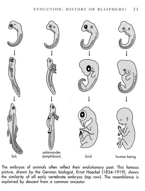
Page 23:
The embryos of animals often reflect their evolutionary past. This famous picture, drawn by the German biologist, Ernst Haeckel (1834-1919), shows the similarity of all early vertebrate embryos (top row). The resemblance is explained by descent from a common ancestor.
[1627] Book: The Discovery of Evolution. By David Young. Cambridge University Press, 1992.
Page 146 shows this drawing, explicitly labeled as Haeckel's:
Page 147:
Haeckel was able to show that at this early stage there was not much to chose between the embryos of bird, dog and human. They all resemble a simplified vertebrate. Only at a later stage do the differences between them make their appearance. Hence the study of embryos provided good evidence for the common ancestry of all vertebrates, including humans. ... But it soon became clear that recapitulation did not hold up to the detailed level Haeckel had hoped for, and his theory lost its appeal after the turn of the century. Most zoologists were content to use embryology as evidence for evolution in general, without expecting it to yield detailed information on phylogeny.
[1628] Book: The Human Body: an Introduction to Structure and Function. By Adolf Faller, Michael Schünke, Gabriele Schünke. Thieme Medical Publishers, 2004. Translated and revised from the 13th German edition (1999) by Oliver French, M.D. Page 60:
[1629] Textbook: Biology. By Peter H. Raven & George B. Johnson. Fifth edition (customized). McGraw-Hill, 1999. Page 416:
The fact that seemingly different organisms exhibit similar embryological forms provides direct evidence of an evolutionary relationship.

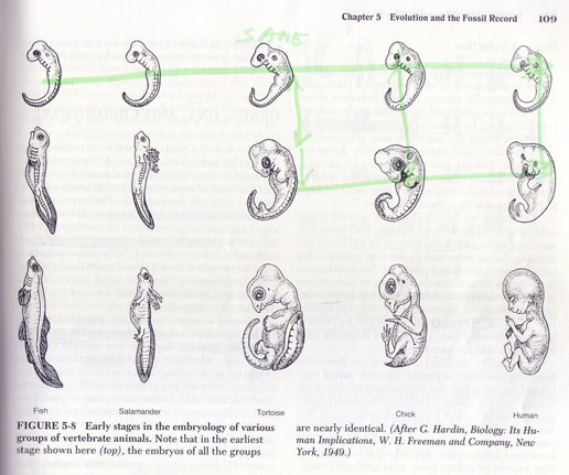
[1630] Book: Exploring Earth and Life Through Time. By Steven M. Stanley (Johns Hopkins University). W.H. Freeman and Company, 1993. Page 108:
When Darwin returned to England and weighed other evidence indicating that one type of organism had evolved from another, he found that certain anatomical relationships seemed to build and especially compelling case. One such piece of evidence was the remarkable similarity of the embryos of all vertebrate animals (Figure 5.8).
Page 109:
[1631] Web page: "Haeckel and his Embryos." By Ken Miller and Joe Levine. Updated November 21, 1997. http://www.millerandlevine.com/km/evol/embryos/Haeckel.html
"However, his drawings nonetheless became the source material for diagrams of comparative embryology in nearly every biology textbook, including ours!"
NOTE: The yolk sac claim on this webpage is debunked on pages 229-230 of Rational Conclusions.
[1632] Article: "Abscheulich! (Atrocious!)" By Stephen J. Gould. Natural History, March 2000. Pages 42-49.
Page 45 quotes the letter from Richardson, which is August 16, 1999.
[1633] Book: The History of Biology: A Survey. By Erik Nordenskiöld. Tudor Publishing, 1946. Translated from the Swedish volume entitled Biologins Historia, 1920-24.
Directly under the heading "Victory of Darwinism," page 522 states: "During the eighties the dispute as to the justification of Darwinism died down.... [I]t was a time when Gegenbaur's and Haeckel's ideas universally prevailed without opposition...."
[1634] Book: The Golden Age of Zoology: Portraits from Memory. By Richard B. Goldschmidt. University of Washington Press, 1956. Pages 34-35:
When I was a high school boy of about sixteen ... I found Haeckel's history of creation one day and read it with burning eyes and soul. It seemed that all the problems of heaven and earth were solved simply and convincingly; there was an answer to every question which troubled the young mind. Evolution was the key to everything.... There was no creation, no God, no heaven and hell, only evolution and the wonderful law of recapitulation which demonstrated the fact of evolution to the most stubborn believer in creation.
Pages 31-40 reveal that Goldschmidt later came to view Haeckel as a demagogue.
Pages 37- 38:
When I first met Haeckel he was already a septuagenarian. When Richard Hertwig brought him to my room I knew at once, from the many pictures I has seen, who the visitor was. ... I showed Haeckel some slides, but his comments showed that his ideas were still those of the naive phylogeny of his youth. The last time I saw Haeckel he was past eighty and in bad shape.
Page 34: "This role he assumed and established in his two major popular books, the Natural History of Creation and Anthropogeny. The present generation can hardly understand the influence Haeckel exercised through these books upon the minds of youth, of laymen in general, and also upon large sections of the professional world."
[1635] Web page: "The Material Basis of Evolution – Reissued." Yale University Press. Accessed September 22, 2007 at http://yalepress.yale.edu/yupbooks/book.asp?isbn=9780300028232
"Goldschmidt, one of the world's great geneticists, delivered the prestigious Silliman lectures at Yale University in 1939 and published his remarks in 1940 as The Material Basis of Evolution."
[1636] Web page: "Haeckel and his Embryos." By Ken Miller and Joe Levine. Updated November 21, 1997. http://www.millerandlevine.com/km/evol/embryos/Haeckel.html
"Our books now contain accurate drawings of the embryos made from detailed photomicrographs."
NOTE: The yolk sac claim is debunked on pages 229-230 of Rational Conclusions.
[1637] Textbook: Biology. By Kenneth R. Miller & Joseph Levine. Prentice Hall, 2000. Page 283.
[1638] Textbook: Biology (Teachers' Edition). By Kenneth R. Miller & Joseph Levine. Pearson Education, 2004. In the section entitled "Evidence of Evolution," page 385 states:
Similarities in Embryology The early stages, or embryos, of many animals with backbones are very similar. This does not mean that a human embryo is ever identical to a fish or a bird embryo. However, as you can see in Figure 15-17, many embryos look especially similar during certain stages of development.
There have, in the past, been incorrect explanations for these similarities. Also, the biologist Ernst Haeckel fudged some his drawings to make the earlier stages of some embryos seem more similar than they actually are! Errors aside, however, it is clear that the same groups of embryonic cells develop in the same order and in similar patterns to produce the tissues and organs of all vertebrates. These common cells and tissues, growing in similar ways, produce the homologous structures discussed earlier.
[1639] For one of many citations containing evidence that blows holes in the citation above:
Paper: "Inverting the hourglass: quantitative evidence against the phylotypic stage in vertebrate development." By Olaf R. P. Bininda-Emonds & others. Proceedings of the Royal Society: Biological Sciences, January 20, 2003. http://www.pubmedcentral.nih.gov/articlerender.fcgi?artid=1691251
Page 344:
Support for the hourglass definition of the phylotypic stage derives largely from subjective statements about the overall similarity of embryos of different species, usually based on an examination of pictures of embryos and not from rigorous character-based data analysis. ...
... Shared features undoubtedly exist during the mid-embryonic period, but those used to support the phylotypic stage are often defined so coarsely as to obscure potential variation between species. For instance, the statement that vertebrate embryos all possess a heart during the phylotypic stage (Kimmel et al. 1995) ignores important variation in how the heart is formed (see Richardson 1995) as well as the existence of heterochrony, which can result Proc. R. Soc. Lond. B (2003) in the heart being in different stages of its development when other key characters are all present (which may themselves be at varying stages in their development).
[1640] For another example:
Paper: "On the law of development commonly known as von Baer's law; and on the significance of ancestral rudiments in embryonic development." By Adam Sedgwick. Quarterly Journal of Microscopical Science, April 1, 1894. Pages 35-52. http://jcs.biologists.org/cgi/reprint/s2-36/141/35
Pages 38-39:
If v. Baer's law has any meaning at all, surely it must imply that animals so closely allied as the fowl and duck would be indistinguishable in the early stages of development ... yet I can distinguish a fowl and a duck embryo on the second day by the inspection of a single transverse section through the trunk.... But it is not necessary to emphasize further these embryonic differences; every embryologist knows that they exist and could bring forward innumerable instances of them. I need only say with regard to them that a species is distinct and distinguishable from its allies from the very earliest stages all through the development, although these embryonic differences do not necessarily implicate the same organs as do the adult differences.
[1641] Book: 5 Steps to a 5: AP Biology. By Mark Anestis. McGraw-Hill, 2002.
Page 133 lists three kinds of evidence that provide "support for the theory of evolution." One of these is described as such:
The study of embryos reveals remarkable similarities between organisms at the earliest stages of life, although as adults (or even at birth) the species look completely different. Human embryos, for example, actually have gills for a short time during early development, hinting at our aquatic ancestry.
[1642] Paper: "On the Respiratory Branchial Apparatus of the Human Embryo during the first three months of its growth." By M. Serres. Compte Rendu des Séances de 1'Academie des Sciences, June 17, 1839. Translated and summarized in the Edinburgh Medical and Surgical Journal. Volume 52, 1839.
Page 567 states that "the fissures in the lateral parts of the neck of the embryo, which M. Rathke discovered in 1825, and which the analogy of the lower animals led him to consider as the respiratory apparatus of the embryo...."
[1643] Paper: "On the Branchial or Gill-like Openings in the Neck of the Human Fetus, as a Cause of Certain Malformations." By M. Ascherson. Translated and summarized in the Dublin Journal of Medical and Chemical Science, Volume 5, 1834. Page 314:
To one of these transition forms belong the branchial fistule discovered by Rathke, first in the young of the pig, horse, hen, water-snake, (coluber natrix,) and lizard, and afterwards in a human embryo, about seven or eight weeks old. These fistule or tubes consist in from six to eight apertures, arranged symmetrically on either side of the neck, opening into the pharynx, covered externally with a sort of operculum, and exhibiting on their inner surface several arched lamella; Rathke compares these apertures with the branchial apertures of the shark....
[1644] Book: A Theoretical and Practical Treatise on Midwifery. By P. Cazeaux. Second American edition translated from the 5th French edition. Lindsay & Blakiston, 1857. Page 230:
If something analogous to respiration in the adult be sought for in the functions of the fetus, this question will doubtless be answered negatively; because the atmospheric air, having no access to it whatever, the fetal blood could not possibly obtain any elements from it. ...
According to some, the liquor amnii [amniotic fluid] is the modifying agent for the blood, and Beclard supposes that the lungs are the seat of such changes, the amniotic liquid reaching them through the air-passages. Agreeably to M. Geoffroy St. Hilaire, the whole surface of the child's body absorbs air, or a vivifying gas, like insects, by a species of air-tubes, or by minute fissures which exist on the lateral parts of the neck in young embryos. The resemblance between those fissures and the branchial apparatus in the fish has given rise to the belief of an analogous function; hence, they are called the branchial fissures.
NOTE: As we shall see below, the author does not accept this claim.
[1645] Textbook: Biology: Investigating Life on Earth. By Vernon L. Avila. Second edition. Jones and Bartlett, 1995.
Page 691: "The exchange of gases, nutrients, and wastes between mother and embryo occurs through the membranes of the chorionic villi."
Page 693: "Small pools of maternal blood surround the chorionic villi. These pools are fed by maternal blood vessels, which connect to the circulatory system of the mother. ... The exchange of nutrients, gases, and wastes between embryo and mother takes place through the placenta."
[1646] Textbook: Fundamentals of Anatomy & Physiology. By Frederic H. Martini (Ph.D. in comparative and functional anatomy from Cornell University) Prentice Hall, 2001.
The text and series of diagrams on pages 1068-72 trace the development of the chorionic villi, placenta, and umbilical cord from the time that the blastocyst (early preborn human) implants in the uterus.
[1647] Paper: "On the Respiratory Branchial Apparatus of the Human Embryo during the first three months of its growth." By M. Serres. Compte Rendu des Séances de 1'Academie des Sciences, June 17, 1839. Translated and summarized in the Edinburgh Medical and Surgical Journal. Volume 52, 1839. Page 567:
M. Serres demonstrates in this paper that the fissures in the lateral parts of the neck of the embryo, which M. Rathke discovered in 1825, and which the analogy of the lower animals led him to consider as the respiratory apparatus of the embryo, do not perform that function; but he proves satisfactorily, that this function is performed by a villous structure, which he has discovered traversing the thickness of the decidua reflexa.... He has arrived at this from numerous dissections, which he narrates at length. ...
... As the ovum increases in size, however, a portion of the villosities of the chorion go to form the placenta, where the fetal respiration is afterwards carried on....
[1648] Article: "Fetus." Black's Medical Dictionary. Edited by Gordon Macpherson. 39th edition. Madison Books, 1999. Pages 202-3.
Page 202: "After fertilization with a spermatozoon the ovum becomes embedded in the mucous membrane of the uterus, its covering being known as the decidua."
[1649] Book: Embryology (Board Review Series). By Ronald W. Dudek & James D. Fix. Second edition. Lippincott Williams & Wilkins, 1998.
The rear cover states that this book is "designed for medical students."
Page 149: "Pharyngeal apparatus. ...contributes greatly to formation of the head and neck."
Pages 150-2 contain details of their histology and outlines what parts of the face and neck each arch becomes.
NOTE: This book does not even mention the outdated and misleading term "branchial."
[1650] Textbook: Before We Are Born: Essentials of Embryology and Birth Defects. By Keith L. Moore & T.V.N. Persaud. Saunders, 2003. Sixth edition.
Page 152: "Because gills do not form in human embryos, the term pharyngeal arch is now used instead of branchial arch. ... The pharyngeal arches contribute extensively to the formation of the face, nasal cavities, mouth, larynx, pharynx, and neck."
[1651] Textbook: Langman's Medical Embryology. By T. W. Sadler. Ninth edition. Lippincott Williams & Wilkins, 2004. Pages 363-4:
The most typical feature in development of the head and neck is formed by the pharyngeal or branchial arches. ... Initially, they consist of bars of mesenchymal tissue separated by deep clefts known as pharyngeal (branchial) clefts (Figs 15.3C; see also 15.6). Simultaneously, with the development of the arches and clefts, a number of outpocketings, the pharyngeal pouches, appear along the lateral wall of the pharyngeal gut.... Although development of pharyngeal arches, clefts and pouches resembles formation of gills in fishes and amphibia*, in the human embryo real gills (branchia) are never formed. Therefore, the term pharyngeal (arches, clefts, and pouches) has been adopted for the human embryo.
Pharyngeal arches not only contribute to the formation of the neck, but also play an important part in formation of the face.
NOTES: As shown on pages 217-218 of Rational Conclusions, this is simply not true. The author (Sadler ) is an authority in human embryos, but not an authority with fish and amphibian embryos. He is still clearly under the impression of Haeckel's fraudulent drawings.
Pages 366-372 explain what develops from each pharyngeal arch.
Pages 372-375 explain what develops from each pharyngeal pouch.
Page 375 explains what develops from the pharyngeal clefts.
[1652] Entry: "branchial." Merriam-Webster's Collegiate Dictionary, Encyclopedia Britannica Ultimate Reference Suite 2004.
[1653] Entries: "pharyngeal, pharynx." Merriam-Webster's Collegiate Dictionary, Encyclopedia Britannica Ultimate Reference Suite 2004.
[1654] Book: On the Origin of Species by Means of Natural Selection, or the Preservation of Favoured Races in the Struggle for Life. By Charles Darwin. John Murray, 1859. http://www.literature.org/authors/darwin-charles/the-origin-of-species/
Preface: "Lamarck was the first man whose conclusions on the subject excited much attention. This justly-celebrated naturalist first published his views in 1801...."
[1655] Book: Zoological Philosophy: An Exposition with Regard to the Natural History of Animals. By J. B. Lamarck. Published in 1809. Translated and introduced by Hugh Elliot. Macmillan and Co, 1914; 1963 reprint by Hafner Publishing.
Page 175: "I do not doubt that mammals originally came from the water, nor that water is the true cradle of the entire animal kingdom."
[1656] Book: Victorian Sensation: The Extraordinary Publication, Reception, and Secret Authorship of Vestiges of the Natural History of Creation. By James A. Secord. University of Chicago Press, 2000. Pages 2-3:
Contemporaries called it the biggest literary phenomenon for decades, perhaps bigger than even Charles Dickens's early novels. The book was mentioned in thousands of letters and diaries, denounced and praised in pulpits ... reviewed in scores of periodical and pamphlets, and in Britain alone sold fourteen editions and almost forty thousand copies.
Page 4: "In archives, newspapers, and memoirs, there are thousands of traces of encounters with the book." The graph on page 526 shows that total sales of the Origin of Species (1859) did not catch up with sales of Vestiges of the Natural History of Creation (1844) until 1882, and "did not decisively overtake Vestiges until the twentieth century."
[1657] Book: On the Origin of Species by Means of Natural Selection, or the Preservation of Favoured Races in the Struggle for Life. By Charles Darwin. John Murray, 1859. http://www.literature.org/authors/darwin-charles/the-origin-of-species/
Preface:
The 'Vestiges of Creation' appeared in 1844. In the tenth and much improved edition (1853).... The work, from its powerful and brilliant style, though displaying in the earlier editions little accurate knowledge and a great want of scientific caution, immediately had a very wide circulation. In my opinion it has done excellent service in this country in calling attention to the subject, in removing prejudice, and in thus preparing the ground for the reception of analogous views.
[1658] Book: Vestiges of the Natural History of Creation. By Anonymous [Robert Chambers]. John Churchill, 1844. Electronic edition prepared by Robert Robbins. http://www.esp.org/books/chambers/vestiges/facsimile/
Page 193: "In mammifers, the gills exist and act at an early stage of the fetal state, but afterwards go back and appear no more; while the lungs are developed."
NOTE: See the next citation revealing that the author continued to promulgate this view in later editions, including the same one that Darwin praised in the citation above.
[1659] Book: Vestiges of the Natural History of Creation. By Anonymous [Robert Chambers]. Tenth edition. John Churchill, 1853.
Page 144: "The mammalia, as creatures destined to breathe the air, are furnished with lungs; but, at an early stage of the fetal progress, this is not the case. They have at that time a branchial apparatus."
[1660] Article: "Sedgwick, Adam." Encyclopedia Britannica Ultimate Reference Suite 2004.
[1661] Book: A Discourse on the Studies of the University of Cambridge. By Adam Sedgwick (Not to be confused with his great nephew of the same name. See citation 1563). Fifth edition. Cambridge University Press, 1850. Page xix:
Is this doctrine true? Has the animal kingdom been first produced by spontaneous generation, and afterwards perfected by transmutation and progressive development? The Author of the Vestiges of the Natural History of Creation has adopted the whole scheme which has been sketched in the preceding sentences; and to a comment on his principles I must devote a portion of this Preface.
Page xxxi: "But during the corresponding period in the gestation of a mammal, no tufts or gills are found in the (so-called) "branchial fissures'" of the embryo. No microscopic power has ever shown the minutest germination of branchial tufts or gills."
[1662] Web page: "Who was Robert Bentley Todd?" King's College, University of London. Accessed September 19, 2007 at http://www.kcl.ac.uk/depsta/iss/archives/175th/faq39.htm
The eminent physiologist, Robert Bentley Todd (1809-1860), was instrumental in setting up King's College Hospital. ... His contributions to medical science were considerable, not least in understanding the physiology of the brain, and owing to his editorship of the seminal, Cyclopaedia of Anatomy and Physiology, published between 1835 and 1859.
[1663] Article: "Sir William Bowman. By Parker Heath. Bulletin of the American Library Association, May 1936. Pages 205-8. http://www.pubmedcentral.nih.gov/picrender.fcgi?artid=234136&blobtype=pdf
Page 206: "With Todd he joined in publishing a work called "Physiological Anatomy and Physiology of Man." ... [I]t is the first physiology work in which histology is given. It is enormously superior to other work in this field and its time."
[1664] Book: The Physiological Anatomy and Physiology of Man. By Robert Bentley Todd & William Bowman. Volume 2. John W. Parker & Son, 1856.
Page 589: "The term branchial arch is a bad one, since it conveys the idea that, at a certain period, branchie are developed in the higher vertebrata, which is not the case."
[1665] Article: "Bischoff, Theodor Ludwig Wilhelm." Biography or Third Division of the English Cyclopedia (Supplement). Edited by Charles Knight. Bradbury, Evans & Company, 1872. Pages 244-5:
From the time when he first attained professorial rank his studies, so far as original research are concerned, have been almost exclusively devoted to the development of the egg, and of the embryo, more especially in the Mammalia. In 1840, the Prussian Royal Academy of Science proposed a prize for the best memoir on the embryology of some mammal, and this stimulated him to prosecute his researches on rabbits with greater vigor; and his memoir ... won the first prize, and was published in 1840. Thus encouraged, he pursued his work, and issued similar treatises on the eggs and embryos of the dog (in 1845), the guinea-pig (in 1852), and of the roe-buck (in 1854). These are also illustrated by numerous plates, and have taken rank amongst the best works of this class, and obtained for him a high reputation. ... His fame, by this time had so spread that many universities had asked him to join their professorial staff, but he declined all such offers until in 1854 he was induced to accept the chair of human anatomy and physiology at Munich. ... [A book of his published in 1867] is fully illustrated and contains a chapter on the Darwinian theory, in which he comes to conclusions opposed to the hypothesis of the descent of man from ... [gorillas, orangutans or chimpanzees]. ... As regards our knowledge of the mammalian egg, Bischoff takes a prominent place among those who established and more fully developed the general facts which had been announced by the pioneers in this line of discovery....
[1666] Book: A Theoretical and Practical Treatise on Midwifery. By P. Cazeaux. Second American edition translated from the 5th French edition. Lindsay & Blakiston, 1857. Page 230:
The resemblance between those fissures and the branchial apparatus in the fish has given rise to the belief of an analogous function; hence, they are called the branchial fissures. But, says Bischoff, in the mammifere and man, these arcs never have an organization justifying in the least the supposition of their being intended for respiration: they never have internal nor external branches; nor do we ever see, as in the branchia, vessels distributed either on their surface or in their interior.
[1667] Book: A System of Midwifery: Including the Diseases of Pregnancy and the Puerperal State. By William Leishman. Third American edition, with additions by John S. Parry. Henry C. Lea, 1879. Page 136:
We need not pause here to discuss exploded theories, as to the source from which oxygen is derived by the fetus. The researches of Bischoff proved that, even in the embryo, respiration by means of the branchial fissures is impossible, and that, in point of fact, these structures have no connection whatever with this function, as was at one time erroneously supposed by Geoffroy Saint-Hilaire and others.
[1668] Book: On the Origin of Species by Means of Natural Selection, or the Preservation of Favoured Races in the Struggle for Life. By Charles Darwin. John Murray, 1859. http://www.literature.org/authors/darwin-charles/the-origin-of-species/
Chapter 13: "Recapitulation and Conclusion":
On the principle of successive variations not always supervening at an early age, and being inherited at a corresponding not early period of life, we can clearly see why the embryos of mammals, birds, reptiles, and fishes should be so closely alike, and should be so unlike the adult forms. We may cease marveling at the embryo of an air-breathing mammal or bird having branchial slits and arteries running in loops, like those in a fish which has to breathe the air dissolved in water, by the aid of well-developed branchia.
NOTE: The glossary of this book defines branchia as: "Gills or organs for respiration in water."
[1669] Book: The History of Creation: Or The Development of the Earth and Its Inhabitants by the Action of Natural Causes. By Ernst Haeckel. Translated by E. Ray Lankester. Volume 1. D. Appleton and Company, 1879. From the fourth German edition of the book entitled Naturliche Schöpfungsgeschichte, 1873. The first edition was published in 1868. Page 307:
Everyone surely knows the gill-arches of fish, those arched bones that lie behind one another ... and which support the gills, the respiratory organs of the fish. ... Now these gills arches originally exist exactly the same in man (D), in dogs (C), in fowls (B), and in tortoises (A), as well as in all other vertebrate animals.
NOTE: The assertions above refer to these drawings, which appear on the unnumbered pages following page 306:
[1670] Book: The Evolution of Man: A Popular Exposition of the Principal Points of Human Ontogeny and Phylogeny. By Ernst Haeckel. Volume 1. D. Appleton and Company, 1896. Translated from the German book entitled Anthropogenie, which was first published in 1874.
Page 360: "In the first stage (upper Row of Section I.), in which the head with the five brain-bladders, and the gill-arches are indeed begun, though the limbs are still entirely wanting, the embryos of all the Vertebrates from Fish to Man differ not at all, or only in non-essential points."
This drawing appears on the unnumbered pages following page 362:
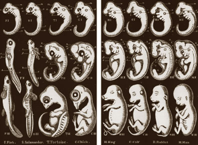
[1671] See citations 1641, 1672, 1676, & 1684.
[1672] Article: "Hawaii's Unearthly Worms." By Jennifer S. Holland. National Geographic, February 2007. Pages 118-131.
Page 123: "An acoel (opposite), a tiny flatworm without a gut ... has a liver (the nubs along its body) and gill slits like those of sharks—and embryonic humans."
[1673] Article: "Evolution." Encyclopedia Britannica Ultimate Reference Suite 2004. In the section entitled "The evidence for evolution."
[1674] Book: The Discovery of Evolution. By David Young. Cambridge University Press, 1992.
Page 146 shows this drawing from Haeckel, explicitly labeled as such:
Page 179: "The gill slits that occur in a mammalian embryo for example, obviously correspond to the gill slits of embryonic fish and not the gills of adult fish."
[1675] Book: The Human Body: an Introduction to Structure and Function. By Adolf Faller, Michael Schünke, Gabriele Schünke. Thieme Medical Publishers, 2004. Translated and revised from the 13th German edition (1999) by Oliver French, M.D. Page 59:
[D]uring development, the germ cell of almost all vertebrates, including humans, go through an embryonic stage in which they bear a remarkable resemblance to a fish embryo. They form gill arches, even though they never develop a complete gill apparatus. This observation serves as evidence that the evolution of vertebrates began with forms that lived in water and breathed through gills.
Page 60:
[1676] Textbook: Biology: Concepts and Connections. By Neil A. Campbell and others. Second edition. Benjamin/Cummings Publishing Company, 1997. Page 264:
[C]omparative embryology ... is another major source of evidence for the common descent of organisms. ... One sign that vertebrates evolved from a common ancestor is that all of them have an embryonic stage in which structures called gill pouches appear on the sides of the throat. At this stage, the embryos of fishes, frogs, snakes, birds, apes – indeed, all vertebrates – look more alike than different.
[1677] Textbook: The Science of Evolution. By William D. Stansfield (Professor of Biological Sciences at California Polytechnic State University). Macmillan, 1977. Page 109:
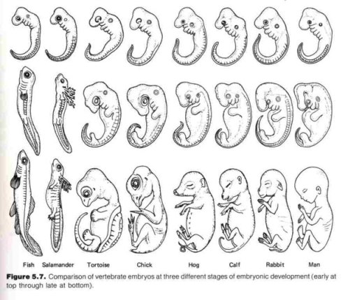
Page 110: "Why should the mammalian embryo have to pass through a stage in which it forms gill arches and gill slits if these structures are never to function as such? The most logical answer is that mammals have retained some genetic information in common with their fish-like ancestors."
[1678] Book: Vertebrate Paleontology and Evolution. By Robert L. Carroll. W.H. Freeman and Company, 1988.
Page 9: "Terrestrial vertebrates go through a stage in which they have fishlike gill slits...."
[1679] Book: Kaplan AP Biology 2005. By Glenn E. Croston. Kaplan Publishing, 2005.
Page 173: "The human embryo develops gill slits, suggesting a relation to the fishes and other vertebrates, but in humans the gills slits evolve later in development and perform other functions."
[1680] Book: Philosophy of Biology. By Elliott Sober. Second edition. Westview Press, 2000.
NOTE: Dr. Sober is a Professor of Philosophy at the University of Wisconsin-Madison. He has authored/edited three books about evolutionary biology that have been published by the MIT Press (Conceptual Issues in Evolutionary Biology, 2006; Reconstructing the Past, 1991; The Nature of Selection, 1985).
Page 40: "Human fetuses develop gill slits and then lose them. ... Gill slits lost their advantage somewhere in the lineage leading to us, so they were deleted from the adult phenotype. Their presence in the embryo did not harm, so the embryonic trait has persisted."
[1681] Book: Fundamentals of Comparative Embryology of the Vertebrates. By Alfred F. Huettner. Revised edition. Macmillan Company, 1949. First edition published in 1941.
Page 40: "When the mammalian embryo develops gill clefts, it is for the same reason that the fish embryo develops them. In the former they appear because the genes for them are still present and have not been changed by natural selection. ... It is in this sense that we can accept the recapitulation theory."
[1682] Book: Evolution and Genetics. By David J. Merrell. Holt, Rinehart and Winston, 1962. Page 90:
The gill arches and the gill slits in the mammalian embryos do not represent the adult ancestral fish, but are similar to those of a fish embryo at a comparable stage of development. ... The obvious question is why there should be a stage in the mammalian embryo where gills and gill arches, which never function as such, are nevertheless present, even though they differentiate into quite different adult structures. The most obvious answer is that the mammals are descended from fishlike ancestors....
[1683] Textbook: Biology. By Peter H. Raven & George B. Johnson. Fifth edition (customized). McGraw-Hill, 1999.
Page 416: "For example, early in their development, human embryos possess gill slits, like a fish...."
[1684] Book: What Evolution Is. By Ernst Mayr. Basic Books, 2001. Page 27:
All of these embryos begin with the same number of gill arches.
Page 30: "[A]ll terrestrial vertebrates (tetrapods) develop gill arches at a certain stage in their ontogeny."
[1685] Book: The Tapir's Morning Bath: Mysteries of the Tropical Rain Forest and the Scientists Who Are Trying to Solve Them. By Elizabeth Royte. Houghton Mifflin, 2001.
Page 183: "At some point in their embryonic development, all terrestrial vertebrates, today and for the last three hundred million years, sported gill arches, just like their marine ancestors."
[1686] Article: "Inside The Womb." By J. Madeleine Nash. Time, November 11, 2002. Pages 68-78.
Page 71: "40 days - At this point, a human embryo looks no different from that of a pig, chick or elephant. All have a tail, a yolk sac and rudimentary gills."
[1687] Book: 5 Steps to a 5: AP Biology. By Mark Anestis. McGraw-Hill, 2002. Page 133:
Support for the theory of evolution can be found in varied kinds of evidence.... The study of embryos reveals remarkable similarities between organisms at the earliest stages of life, although as adults (or even at birth) the species look completely different. Human embryos, for example, actually have gills for a short time during early development, hinting at our aquatic ancestry. ... Darwin used embryology as an important piece of evidence for the process of evolution.
[1688] Textbook: Asking About Life. By Allan J. Tobin & Jennie Dusheck. Third Edition. Brooks Cole, 2004.
Page 316: "The early embryos of vertebrates are amazingly alike (Figure 15 -14). For example, all vertebrate embryos, including humans, have tails and gill-like branchial arches."
[1689] Book: Growth and Development. By Virginia B. Silverstein, Alvin Silverstein & Laura Silverstein Nunn. Twenty-First Century Books, 2008.
Page 69: "All vertebrates look like one another in certain stages of their early development. A mammal goes through fishlike and reptilelike stages during its development before birth. For example, fish, turtles, chickens, mice and humans all develop fishlike tails and gills during the embryo phase."
[1690] Textbook: Evolution. By Monroe W. Strickberger. Third edition. Jones and Bartlett, 2000.
Page 44: Beneath a drawing derived from Haeckel's (explicitly labeled as such), the author states: "Note that each of the embryos begins with a similar number of pharyngeal (gill) arches (pouches below the head) and a similar vertebral column."
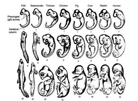
[1691] Textbook: Before We Are Born: Essentials of Embryology and Birth Defects. By Keith L. Moore & T.V.N. Persaud. Saunders, 2003. Sixth edition. Page 152.
[1692] Textbook: Langman's Medical Embryology. By T. W. Sadler. Ninth edition. Lippincott Williams & Wilkins, 2004. Page 364.
[1693] Book: Shaum's Outlines: Biology. By George H. Fried & George J. Hademenos. Second edition, 1999. Page 348.
NOTE: The cover states this book is "Ideal preparation for the MCAT."
[1694] Article: "Evolution." Contributor: Alan R. Templeton (Ph.D., Charles Rebstock Professor of Biology and Professor of Genetics and Biomedical Engineering, Washington University). World Book Encyclopedia, 2007 Deluxe Edition.
[1695] Textbook: Biology. By Peter H. Raven & George B. Johnson. Fifth edition (customized). McGraw-Hill, 1999. Page 417:
Many organisms possess vestigial structures that have no apparent function, but that resemble structures their presumed ancestors had. ... It is difficult to understand vestigial structures such as these as anything other than evolutionary relicts, holdovers from the evolutionary past. They argue strongly for the common ancestry of the members of the groups that share them, regardless of how different they have subsequently become.
[1696] Book: Biology Builder for Standardized Tests. By James R Ogden. Research & Education Association, 1998.
Page 4: "If you are preparing to take the AP Biology, ASVAB, Clep General Biology, GRE Biology, MCAT ... you will be taking a test that requires excellent knowledge of biology. This book present a comprehensive biology review that can be tailored to your specific test preparation needs."
Page 216: "PROBLEM: Describe the various types of evidence from living organisms which support the theory of evolution. SOLUTION: ... the presence of vestigial organs, which are useless or degenerate structures found in the body, points to the existence of some ancestral forms in which these organs were once functional."
[1697] Textbook: The Science of Evolution. By William D. Stansfield (Professor of Biological Sciences at California Polytechnic State University). Macmillan, 1977.
Page 121: "Probably one of the most dramatic lines of evidence for evolution is seen in vestigial or rudimentary structures. These nonfunctional structures and organs are easily explained by the theory of evolution as the now useless remnants of functional structures in ancestral stock."
[1698] Book: Great Ideas of Science: Evolution. By Paul Fleischer. Twenty-First Century Books, 2006.
Page 40: Body structures don't always make sense. ... Many creatures have vestigial organs, for example. These body parts no longer serve a purpose. Vestigial organs are leftovers from a species' evolutionary history."
NOTE: The publisher's website (http://lernerbooks.com) states the reading level is Grade 8 and the interest level is Grades 9-12.
[1699] Book: The Evolutionary Process: A Critical Review of Evolutionary Theory. By Verne Grant (Ph.D. in botany and genetics from Berkeley, Professor of Botany at The University of Texas at Austin). Columbia University Press, 1985.
Page 12: "Vestigial organs. Some members of a major group often possess an organ that is atrophied and non-functional. ... There is no good explanation for the existence of useless rudimentary organs in the doctrine of creationism."
[1700] Book: Exploring Earth and Life Through Time. By Steven M. Stanley (Johns Hopkins University). W.H. Freeman and Company, 1993.
Page 108: "The existence of vestigial organs – organs that serve no apparent purpose but resemble organs that do perform functions in other creatures – further supported Darwin's argument in favor of evolution."
[1701] Book: Zoological Philosophy: An Exposition with Regard to the Natural History of Animals. By J. B. Lamarck. Published in 1809. Translated and introduced by Hugh Elliot. Macmillan and Co, 1914; 1963 reprint by Hafner Publishing. Page 108:
[N]ew needs which establish a necessity for some part really bring about the existence of that part, as a result of efforts; and that subsequently its continued use gradually strengthens, develops and finally greatly enlarges it; in the second place, we shall see that in some cases, when the new environment and new needs have altogether destroyed the utility of some part, the total disuse of that part has resulted in its gradually ceasing to share in the other parts of the animal; it shrinks and wastes little by little, and ultimately, when there has been total disuse for a long period, the part in question ends by disappearing. All this is positive; I propose to furnish the most convincing proofs of it.
[1702] Book: On the Origin of Species by Means of Natural Selection, or the Preservation of Favoured Races in the Struggle for Life. By Charles Darwin. John Murray, 1859. http://www.literature.org/authors/darwin-charles/the-origin-of-species/
Chapter 13: "Mutual Affinities of Organic Beings: Morphology: Embryology: Rudimentary Organs":
I have now given the leading facts with respect to rudimentary organs. In reflecting on them, every one must be struck with astonishment: for the same reasoning power which tells us plainly that most parts and organs are exquisitely adapted for certain purposes, tells us with equal plainness that these rudimentary or atrophied organs, are imperfect and useless. ...
On my view of descent with modification, the origin of rudimentary organs is simple. ...I doubt whether species under nature ever undergo abrupt changes. I believe that disuse has been the main agency; that it has led in successive generations to the gradual reduction of various organs, until they have become rudimentary, as in the case of the eyes of animals inhabiting dark caverns, and of the wings of birds inhabiting oceanic islands, which have seldom been forced to take flight, and have ultimately lost the power of flying. ...
... On the view of descent with modification, we may conclude that the existence of organs in a rudimentary, imperfect, and useless condition, or quite aborted, far from presenting a strange difficulty, as they assuredly do on the ordinary doctrine of creation, might even have been anticipated, and can be accounted for by the laws of inheritance.
[1703] Book: Studies on Fermentation: The Diseases of Beer, Their Causes, and The Means of Preventing Them. By Louis Pasteur. Translated with the author's sanction by Frank Faulkner & D. Constable Robb. Macmillan & Co., 1879. Kraus Reprint Co., 1969. Page 42:
When we see beer and wine undergo radical changes, in consequence of the harbor which those liquids afford to microscopic organisms that introduce themselves invisibly and unsought into it, and swarm subsequently therein, how can we help imagining that similar changes may and do take place in the case of man and animals? Should we, however, be disposed to think that such a thing must hold true, because it seems both probable and possible, we must, before asserting our belief, recall to mind the epigraph of this work: the greatest aberration of the mind is to believe a thing to be, because we desire it.
[1704] Article: "Physiology." Encyclopedia Britannica Ultimate Reference Suite 2004.
The section entitled "Historical background" states: "Physiology as a distinct discipline utilizing chemical, physical, and anatomical methods began to develop in the 19th century."
[1705] Book: Darwiniana. By Thomas H. Huxley. D. Appleton and Company, 1894.
Chapter 4: "The Genealogy of Animals."* Page 114:
Thus, there can be little doubt that the mammary gland was as apparently useless in the remotest male mammalian ancestor of man as in living men, and yet it has not disappeared.† Is it then still profitable to the male organism to retain it? Possibly; but in that case its dysteleological value is gone.1 ...
1 The recent discovery of the important part played by the Thyroid gland should be a warning to all speculators about useless organs. 1893
NOTES:
* This essay was written by Huxley in 1869. The footnote was added by him in 1893.
† See page 230 in Rational Conclusions for discussion of male nipples.
[1706] Article: "Huxley, T.H." Encyclopedia Britannica Ultimate Reference Suite 2004.
[1707] Book: The Evolution of Man: A Popular Exposition of the Principal Points of Human Ontogeny and Phylogeny. By Ernst Haeckel. Volume 2. D. Appleton and Company, 1896. Translated from the German book entitled Anthropogenie, which was first published in 1874. Pages 336-7:
We must say a few words about an interesting rudimentary organ of the respiratory intestine, the thyroid gland (thyreoidea), the large gland situated in front of the larynx, and below the so-called "Adam's apple," and which, especially in the male sex, is often very prominent; it is produced in the embryo by the separation of the lower wall of the throat (pharynx). This thyroid gland is of no use whatever to man; it is only aesthetically interesting, because in certain mountainous districts it has a tendency to enlarge, and in that case it forms the "goitre" which hangs from the neck in front. Its dysteleological interest is, however, far higher; for as Wilhelm Muller of Jena has shown, this useless and unsightly organ is the last remnant of the "hypobranchial groove," which we have already considered....
[1708] Book: The Descent of Man, And Selection in Relation to Sex. By Charles Darwin. Second Edition, John Murray, 1874. 1890 Reprint. First published in 1871.
Page 21: "Not only is it [the human appendix] useless, but it is sometimes the cause of death...."
[1709] Book: Shaum's Outlines: Biology. By George H. Fried & George J. Hademenos. Second edition, 1999.
Page 348: "It would be much harder to account for nonfunctional vestiges such as the human appendix or coccyx (a remnant of tail vertebrae), if each creature were created by divine design."
NOTE: The cover states this book is "Ideal preparation for the MCAT."
[1710] Book: 5 Steps to a 5: AP Biology. By Mark Anestis. McGraw-Hill, 2002. Pages 133-4:
Support for the theory of evolution can be found in varied kinds of evidence: ... [The author provides three examples, the third of which is:] Vestigial characters. Most organisms carry characters that are no longer useful, although they once were. ... Darwin used vestigial characters as evidence in his original formulation of the process of evolution, listing the human appendix as an example.
[1711] Textbook: Biology. By Kenneth R. Miller & Joseph Levine. Prentice Hall, 1993.
Page 284: "This appendix does not seem to serve a useful purpose today."
[1712] Book: The New Dictionary of Cultural Literacy. By E. D. Hirsch, Joseph F. Kett, & James S. Trefil. Houghton Mifflin Books, 2002.
Page 549: "The appendix has no known function in present-day humans, but it may have played a role in the DIGESTIVE SYSTEM in humans of earlier times."
[1713] Book: Kaplan AP Biology 2005. By Glenn E. Croston. Kaplan Publishing, 2005.
Page 173: "The appendix, small and useless in humans, assists digestion of cellulose of herbivores...."
[1714] Book: Finding Darwin's God. By Kenneth R. Miller. Cliff Street Books, 1999.
Page 101: "Our appendix, for example, seems to serve only to make us sick...."
[1715] Textbook: Surgery: Basic Science and Clinical Evidence. Edited by Jeffrey A Norton, R. Randal Bollinger, Alfred E. Chang, Stephen F. Lowry, Sean J. Mulvihill, Harvey I. Pass, Robert W. Thompson. Springer, 2001. Section entitled "Appendix." By David J. Soybel, Department of Surgery, West Roxbury VA Medical Center. Page 649:
With regard to function, the widely held notion that the appendix is a vestigial organ is not consistent with the facts. Curiously, the appendix seems more highly developed in the higher primates, arguing against a vestigial role. Recent studies have focused on characterizing immune cell populations and their response to luminal antigens, offering the possibility that the appendix may play a role in immune surveillance. Although the unique function of the appendix remains unclear, the mucosa of the appendix, like any other mucosal layer, is capable of secreting fluid, mucin, and proteolytic enzymes.3
NOTE: Pages xvii-xxiv contains a list of 140 M.D.s/Ph.D.s who contributed to this work. The Preface states that this book is the result of a "dream" to "assemble the current and future leaders in surgery and ask them to develop an evidenced based surgical textbook that would provide the reader with the most up-to-date and relevant information on which to base decisions in modern surgical practice. In other words, the dream was to create the best, most comprehensive textbook of surgery."
[1716] Web page: "Ask the Experts: What is the function of the human appendix? Did it once have a purpose that has since been lost?" Answered by Loren G. Martin (Professor of Physiology at Oklahoma State University). Scientific American (online), October 21, 1999. http://www.scientificamerican.com/article.cfm?...
[1717] Textbook: Fundamentals of Anatomy & Physiology. By Frederic H. Martini (Ph.D. in comparative and functional anatomy from Cornell University). Prentice Hall, 2001.
Page 9: [The lymphatic system:] "Defends against infection and disease; returns tissue fluid to the bloodstream." [More details on page 752.]
Page 882: "The cecum collects and stores material from the ileum and begins the process of compaction. ... The mucosa and submucosa of the appendix are dominated by lymphoid nodules, and the primary function of the appendix is as an organ of the lymphatic system."
[1718] Book: The Human Body: an Introduction to Structure and Function. By Adolf Faller, Michael Schünke, Gabriele Schünke. Thieme, 2004. Translated and revised from the 13th German edition (1999) by Oliver French.
Page 414: "The appendix ... has an important function in the human specific immune system (see above) [page 296]."
Page 296:
Because of their large surface area, the intestines play a central role in immunity. After all, 70-80% of all antibody-producing cells are situated in the intestinal wall. ... Diffuse collections and loose associations of lymphocytes (lymphatic follicles) can be found throughout the gastrointestinal tract, which because of its direct contact with ingested nutrients, is an ideal portal of entry for antigens [toxic substances]. Organized lymphatic tissue is present in the vermiform appendix.... Into the epithelium [lining] of the intestinal mucosa are dispersed specific cells that apparently selectively recognize and take up antigenic substances.
[1719] Book: Medical Primatology: History, Biological Foundations and Applications. By Eman P. Fridman. Edited by Ronald D. Nadler. Taylor and Francis, 2002. Page 180:
I cannot avoid mentioning the persistent mistake made by scientists who used nonprimates in research, which led to an incorrect conception about the appendix. For many decades, on the basis of using different animals, the vermiform process of the cecum [appendix] was considered a rudimentary organ which lost its function in humans. Only the studies carried out on primates (the appendix appears only in certain monkeys of the Old World, and reaches complete homology with humans in the apes) enabled researches to discover that the appendix is not rudimentary. On the contrary, it is a phylogenetically new structure with active lymphoid, secretory, and incretory functions. It is associated with the bacteriology of the intestines, the activity of the large intestine, and it plays an important role in the immunological process of the organism.
[1720] Paper: "Biofilms in the large bowel suggest an apparent function of the human vermiform appendix." By R. Randal Bollinger and others. Journal of Theoretical Biology, December 21, 2007. Pages 826-831. http://www.ncbi.nlm.nih.gov/pubmed/17936308
Based (a) on a recently acquired understanding of immune-mediated biofilm formation by commensal bacteria in the mammalian gut, (b) on biofilm distribution in the large bowel, (c) the association of lymphoid tissue with the appendix, (d) the potential for biofilms to protect and support colonization by commensal bacteria, and (e) on the architecture of the human bowel, we propose that the human appendix is well suited as a "safe house" for commensal bacteria, providing support for bacterial growth and potentially facilitating re-inoculation of the colon in the event that the contents of the intestinal tract are purged following exposure to a pathogen.
[1721] Article: "Appendix May Have A Purpose After All." Associated Press, October 5, 2007. http://www.cbsnews.com/stories/2007/10/05/health/main3338152.shtml
NOTE: The professor quoted had nothing to do with the study and is a known proselytizer for evolution.
[1722] Book: Histology: A Text and Atlas With Correlated Cell and Molecular Biology. By Michael H. Ross & Wojciech Pawlina. Fifth edition. Lippincott Williams & Wilkins, 2006.
Page 414-5: "With age, the amount of lymphatic tissue within the organ regresses and is difficult to recognize."
[1723] Web page: "Ask the Experts: What is the function of the human appendix? Did it once have a purpose that has since been lost?" Answered by Loren G. Martin (Professor of Physiology at Oklahoma State University). Scientific American (online), October 21, 1999.
http://www.scientificamerican.com/article.cfm?id=what-is-the-function-of-t
[1724] Tutorial: "Histology of the GI Tract." By S. N. Lawson. Bristol Biomedical Image Archive (University of Bristol), 2002. http://www.bristol.ac.uk/phys-pharm/teaching/...
Page 35: "At what age is the maximum amount of follicular lymphoid tissue present in the normal appendix? It increases up to about 10 years and then declines. In the adult there are scattered lymph follicles in the normal appendix."
NOTES: In the previous citation, it is stated that the amount of lymphoid tissue peaks between twenty and thirty years of age. I have been unable to definitively ascertain which of these assertions is more accurate. Pages 29-40 have some exceptional pictures of the appendix.
[1725] Paper: "Biofilms in the large bowel suggest an apparent function of the human vermiform appendix." By R. Randal Bollinger and others. Journal of Theoretical Biology, December 21, 2007. Pages 826-831. http://www.ncbi.nlm.nih.gov/pubmed/17936308
"Further, it is anticipated that the biological function of the appendix may be observed only under conditions in which modern medical care and sanitation practices are absent, adding difficulty to any potential studies aimed at demonstrating directly the role of the appendix in humans."
[1726] Book: The Human Body: an Introduction to Structure and Function. By Adolf Faller, Michael Schünke, Gabriele Schünke. Thieme, 2004. Translated and revised from the 13th German edition (1999) by Oliver French, M.D.
Page 414: "The appendix ... has an important function in the human specific immune system (see above) [page 296]."
Page 296:
Because of their large surface area, the intestines play a central role in immunity. After all, 70-80% of all antibody-producing cells are situated in the intestinal wall. ... Diffuse collections and loose associations of lymphocytes (lymphatic follicles) can be found throughout the gastrointestinal tract, which because of its direct contact with ingested nutrients, is an ideal portal of entry for antigens [toxic substances]. Organized lymphatic tissue is present in the vermiform appendix.... Into the epithelium [lining] of the intestinal mucosa are dispersed specific cells that apparently selectively recognize and take up antigenic substances.
NOTE: Modern farming technologies, rapid distribution capabilities, cleanliness standards, refrigeration, and comforts we take for granted (such as indoor plumbing) greatly limit the amount of antigens in our food.
[1727] Book: Gastroenterology 3: Large Intestine. Edited by John Alexander-Williams & Henry J. Binder. Butterworth's International Medical Reviews (BIMR), 1983. Chapter 1: "Fluid and electrolyte transport in the colon." By P.C. Hawker and L.A. Turnberg. Page 1:
The major functions of the human colon are the absorption of salt and water, and the storage of dehydrated luminal contents until they can be evacuated. There are large concentration gradients for electrolytes across the colonic mucosa and only small amounts of electrolytes escape in normal stools. The normal daily loss in the stool, of less than 5 mmol (mEq) of sodium chloride, helps explain man's ability to survive for long periods despite a very low salt intake. Despite this important role of sodium retention the apparent good health of subjects after total colectomy emphasizes that under normal circumstances a colon is not an essential organ. Nevertheless, some studies suggest that many patients after colectomy have evidence of sodium and possibly potassium depletion, although they are clinically normal.
Page 11: "Far from being an inactive organ of storage the colon has an important role in gastrointestinal fluid and electrolyte conservation."
[1728] Paper: "Appendectomy During Childhood and Adolescence and the Subsequent Risk of Cancer in Sweden." By Judith U. Cope. Pediatrics, June 6, 2003. Pages 1343-1350. http://pediatrics.aappublications.org/cgi/content/full/111/6/1343
A 55% elevated risk for non-Hodgkin's lymphoma (NHL; 25 cases; SIR: 1.55; 95% CI: 1.0–2.3) reflected a significant 73% increase among children who underwent appendectomy with appendicitis and a nonsignificant 34% increase in those who underwent appendectomy without appendicitis. The excess of NHL in both major groups was balanced by a 32% reduced risk for total leukemia (SIR: 0.68; 95% CI: 0.4–1.1). ...
... Risks were >2.4-fold and significantly increased for total stomach cancers (SIR: 2.45; 95% CI: 1.1–4.8) based on 8 cases, all adenocarcinomas. A 3-fold increase in cancers occurring in the eye was observed (5 cases with different histologic types, SIR: 3.03; 95% CI: 0.98–7.1). Risks were significantly decreased for colon cancer (SIR: 0.42; 95% CI: 0.2–0.9). ...
Conclusions. It is reassuring that there was no overall increase of cancer several years after childhood appendectomy. Increased risks for NHL and stomach cancer, occurring 15 or more years after appendectomy, were based on small absolute numbers of excess cancers. As 95% of the subjects were younger than 40 years at exit, this cohort requires continuing follow-up and monitoring.
[1729] Book: Finding Darwin's God. By Kenneth R. Miller. Cliff Street Books, 1999.
Page 101: "Our appendix, for example, seems to serve only to make us sick...."
NOTE: Observe from citation 1716 that this view was outdated at the time Miller voiced it.
[1730] Article: "The Progress of Medicine," By Arthur J. Snider. Science Digest, June 1966. Pages 31-2:
The lowly human appendix, long a source of debate and curiosity as to its importance to health, may be a protector against cancer, according to Dr. Howard R. Bierman, clinical professor of medicine, Loma Linda University School of Medicine, Santa Barbara, Cal.
He has found among several hundred patients with leukemia, Hodgkin's disease, cancer of the colon and cancer of the ovaries that 84 percent had their appendix removed years earlier. In a comparative group without cancer, 75 percent still retained the vestigial organ.
The human appendix may be an immunological organ whose premature removal during its functioning period permits leukemia and other related forms of cancer to begin their development," Dr. Bierman submitted. "The appendix is composed of lymphoid tissue, suggesting that, like other such lymphoid organs as the tonsils and spleen, it may secrete antibodies which protect the body against attacking viral agents."
"Ironically, most of the patients in our study had developed cancer after the 'routine' removal of a perfectly healthy appendix," Dr. Bierman reports. "The operation usually was performed incidentally at the time of some other surgical procedure when the patient was, on the average, 27 years old."
[1731] Textbook: The Science of Evolution. By William D. Stansfield. Macmillan, 1977. Page 123: "The best know [vestigial organ] is the vermiform appendix...."
[1732] Book: The Descent of Man, And Selection in Relation to Sex. By Charles Darwin. Second Edition, John Murray, 1874. 1890 Reprint. First published in 1871. Chapter 1: "The evidence of the descent of man from some lower form":
The sense of smell is of the highest importance to the greater number of mammals—to some, as the ruminants, in warning them of danger; to others, as the Carnivora, in finding their prey; to others, again, as the wild boar, for both purposes combined. But the sense of smell is of extremely slight service, if any, even to the dark colored races of men, in whom it is much more highly developed than in the white and civilized races. (36. The account given by Humboldt of the power of smell possessed by the natives of South America is well known, and has been confirmed by others. M. Houzeau ('Etudes sur les Facultes Mentales,' etc., tom. i. 1872, p. 91) asserts that he repeatedly made experiments, and proved that Negroes and Indians could recognize persons in the dark by their odor. Dr. W. Ogle has made some curious observations on the connection between the power of smell and the coloring matter of the mucous membrane of the olfactory region as well as of the skin of the body. I have, therefore, spoken in the text of the dark-colored races having a finer sense of smell than the white races. See his paper, 'Medico-Chirurgical Transactions,' London, vol. liii. 1870, p. 276.) Nevertheless it does not warn them of danger, nor guide them to their food; nor does it prevent the Esquimaux [Eskimo] from sleeping in the most fetid atmosphere, nor many savages from eating half-putrid meat. In Europeans the power differs greatly in different individuals, as I am assured by an eminent naturalist who possesses this sense highly developed, and who has attended to the subject. Those who believe in the principle of gradual evolution, will not readily admit that the sense of smell in its present state was originally acquired by man, as he now exists. He inherits the power in an enfeebled and so far rudimentary condition, from some early progenitor, to whom it was highly serviceable, and by whom it was continually used. In those animals which have this sense highly developed, such as dogs and horses, the recollection of persons and of places is strongly associated with their odor; and we can thus perhaps understand how it is, as Dr. Maudsley has truly remarked (37. 'The Physiology and Pathology of Mind,' 2nd ed. 1868, p. 134.), that the sense of smell in man "is singularly effective in recalling vividly the ideas and images of forgotten scenes and places."
[1733] Book: Memmler's Structure and Function of the Human Body. By Barbara Janson Cohen. Eighth edition, Lippincott Williams & Wilkins, 2005.
Page 189: "The importance of the sense of smell, or olfaction ... is often underestimated. This sense helps to detect gases and other harmful substances in the environment and helps to warn of spoiled food. Smells can trigger memories and other physiological responses. Smell is also important in sexual behavior."
[1734] Book: Zoological Philosophy: An Exposition with Regard to the Natural History of Animals. By J. B. Lamarck. Published in 1809. Translated and introduced by Hugh Elliot. Macmillan and Co, 1914; 1963 reprint by Hafner Publishing. Page 108:
[N]ew needs which establish a necessity for some part really bring about the existence of that part, as a result of efforts; and that subsequently its continued use gradually strengthens, develops and finally greatly enlarges it; in the second place, we shall see that in some cases, when the new environment and new needs have altogether destroyed the utility of some part, the total disuse of that part has resulted in its gradually ceasing to share in the other parts of the animal; it shrinks and wastes little by little, and ultimately, when there has been total disuse for a long period, the part in question ends by disappearing. All this is positive; I propose to furnish the most convincing proofs of it.
[1735] Book: On the Origin of Species by Means of Natural Selection, or the Preservation of Favoured Races in the Struggle for Life. By Charles Darwin. John Murray, 1859. http://www.literature.org/authors/darwin-charles/the-origin-of-species/
Chapter 13: "Mutual Affinities of Organic Beings: Morphology: Embryology: Rudimentary Organs":
On my view of descent with modification, the origin of rudimentary organs is simple. ...I doubt whether species under nature ever undergo abrupt changes. I believe that disuse has been the main agency; that it has led in successive generations to the gradual reduction of various organs, until they have become rudimentary, as in the case of the eyes of animals inhabiting dark caverns, and of the wings of birds inhabiting oceanic islands, which have seldom been forced to take flight, and have ultimately lost the power of flying. Again, an organ useful under certain conditions, might become injurious under others, as with the wings of beetles living on small and exposed islands; and in this case natural selection would continue slowly to reduce the organ, until it was rendered harmless and rudimentary.
Any change in function, which can be effected by insensibly small steps, is within the power of natural selection; so that an organ rendered, during changed habits of life, useless or injurious for one purpose, might easily be modified and used for another purpose. Or an organ might be retained for one alone of its former functions. An organ, when rendered useless, may well be variable, for its variations cannot be checked by natural selection. At whatever period of life disuse or selection reduces an organ, and this will generally be when the being has come to maturity and to its full powers of action, the principle of inheritance at corresponding ages will reproduce the organ in its reduced state at the same age, and consequently will seldom affect or reduce it in the embryo. Thus we can understand the greater relative size of rudimentary organs in the embryo, and their lesser relative size in the adult. But if each step of the process of reduction were to be inherited, not at the corresponding age, but at an extremely early period of life (as we have good reason to believe to be possible) the rudimentary part would tend to be wholly lost, and we should have a case of complete abortion. The principle, also, of economy, explained in a former chapter, by which the materials forming any part or structure, if not useful to the possessor, will be saved as far as is possible, will probably often come into play; and this will tend to cause the entire obliteration of a rudimentary organ.
As the presence of rudimentary organs is thus due to the tendency in every part of the organization, which has long existed, to be inherited we can understand, on the genealogical view of classification, how it is that systematists have found rudimentary parts as useful as, or even sometimes more useful than, parts of high physiological importance. Rudimentary organs may be compared with the letters in a word, still retained in the spelling, but become useless in the pronunciation, but which serve as a clue in seeking for its derivation. On the view of descent with modification, we may conclude that the existence of organs in a rudimentary, imperfect, and useless condition, or quite aborted, far from presenting a strange difficulty, as they assuredly do on the ordinary doctrine of creation, might even have been anticipated, and can be accounted for by the laws of inheritance.
[1736] Book: Shaum's Outlines: Biology. By George H. Fried & George J. Hademenos. Second edition, 1999.
Page 348: "It would be much harder to account for nonfunctional vestiges ... if each creature were created by divine design."
NOTE: The cover states this book is "Ideal preparation for the MCAT."
[1737] Book: What Evolution Is. By Ernst Mayr. Basic Books, 2001.
Page 31: "[V]estigial structures ... raise insurmountable difficulties for a creationist explanation, but are fully compatible with an evolutionary explanation...."
[1738] Book: On the Origin of Species by Means of Natural Selection, or the Preservation of Favoured Races in the Struggle for Life. By Charles Darwin. John Murray, 1859. http://www.literature.org/authors/darwin-charles/the-origin-of-species/
Chapter 5: "Laws of Variation."
[1739] Article: "Top 10 Useless Limbs (and Other Vestigial Organs)." By Brandon Miller. LiveScience, 2007. http://www.livescience.com/animals/top10_vestigial_organs.html
"Vestigial organs have demonstrated remarkably how species are related to one another, and has given solid ground for the idea of common descent to stand on. ... It is only because of macro-evolutionary theory, or evolution that takes place over very long periods of time, that these vestiges appear."
NOTE: Following the above is a list of ten organs. Number 6 is entitled "The Blind Fish Astyanax Mexicanus." This page states that sighted "fish of the same species live right above, near the surface, where there is plenty of light." How this could be construed as evidence of evolution is beyond me. It is the equivalent of asserting that because a person is born blind, his or her ancestors must have been something other than human.
[1740] Article: Was Darwin Wrong? By David Quammen. National Geographic, November 2004 (cover story). http://ngm.nationalgeographic.com/ngm/0411/feature1/fulltext.html
Vestigial characteristics are still another form of morphological evidence [for evolution], illuminating to contemplate because they show that the living world is full of small, tolerable imperfections. Why do certain species of flightless beetle have wings, sealed beneath wing covers that never open? Darwin raised all these questions, and answered them, in The Origin of Species. Vestigial structures stand as remnants of the evolutionary history of a lineage.
[1741] Genesis 1:31: "And God saw every thing that he had made, and, behold, it was very good. And the evening and the morning were the sixth day."
Genesis 2:16-17: "And the LORD God commanded the man, saying, Of every tree of the garden thou mayest freely eat: But of the tree of the knowledge of good and evil, thou shalt not eat of it: for in the day that thou eatest thereof thou shalt surely die."
Genesis 3: 17-19: "And unto Adam he said, Because thou hast hearkened unto the voice of thy wife, and hast eaten of the tree, of which I commanded thee, saying, Thou shalt not eat of it: cursed is the ground for thy sake; in sorrow shalt thou eat of it all the days of thy life; Thorns also and thistles shall it bring forth to thee; and thou shalt eat the herb of the field; In the sweat of thy face shalt thou eat bread, till thou return unto the ground; for out of it wast thou taken: for dust thou art, and unto dust shalt thou return."
Romans 8:22: "For we know that the whole creation groaneth and travaileth in pain together until now."
[1742] By the way, "the Fall" did not come by man eating an apple as is commonly portrayed. The Bible only refers to the forbidden food as fruit from "the tree of the knowledge of good and evil." Genesis 3:3: "But of the fruit of the tree which [is] in the midst of the garden, God hath said, Ye shall not eat of it, neither shall ye touch it, lest ye die."
The word "fruit" is translated from the Hebrew word "pĕriy," which has three possible meanings: (a) fruit, produce (of the ground), (b) fruit, offspring, children, progeny (of the womb), (c) fruit (of actions). [Entry: "pĕriy (Strong's 06529)." Blue Letter Bible, October 30, 2007.
http://cf.blueletterbible.org/lang/lexicon/lexicon.cfm?...]
[1743] Paper: "Biofilms in the large bowel suggest an apparent function of the human vermiform appendix." By R. Randal Bollinger and others. Journal of Theoretical Biology, December 21, 2007. Pages 826-831. http://www.ncbi.nlm.nih.gov/pubmed/17936308
"In as much as 6 % of the population in industrialized countries, the appendix becomes inflamed and must be surgically removed to avoid a potentially life-threatening infection."
[1744] Abstract: "A murine model of appendicitis and the impact of inflammation on appendiceal lymphocyte constituents." By W. S. Watson Ng and others. Clinical & Experimental Immunology, October 2007. http://www.blackwell-synergy.com/doi/abs/10.1111/j.1365-2249.2007.03463.x
"Data indicate that appendicectomy for intra-abdominal inflammation protects against inflammatory bowel disease (IBD). This suggests an important role for the appendix in mucosal immunity."
[1745] Book: Finding Darwin's God. By Kenneth R. Miller. Cliff Street Books, 1999. Page 101.
[1746] Book: The Encyclopedia of Genetic Disorders and Birth Defects. By James Wynbrandt & Mark D. Ludman. Facts on File, 1991. Preface:
Genetic disorders and birth defects comprise a vast galaxy of anomalous conditions and exert and extraordinary impact on the human population. Attempting even a partial catalog of them is daunting, indeed. More than 4,300 "single gene" disorders have been reported, and are estimated to affect 1% of the population. The number of "multifactorial" disorders, those resulting from a combination of genes, is considered much greater. If late onset disorders are included, 60% of the population is thought to have a genetically influenced disease.
[1747] Book: Population and Evolutionary Genetics: A Primer. By Francisco J. Ayala. Benjamin Cummings Publishing Company, 1982.
Page 24: "Although mutation rates are low, new mutants appear continuously in nature. This is because there are many individuals in any species and many gene loci in each individual."
[1748] Textbook: Principles of Genetics. By D. Peter Snustad & Michael J. Simmons. John Wiley & Sons, 2006. Fourth edition.
Page 348: "Most of the thousands of mutations that have been identified and studied by geneticists are deleterious and recessive."
[1749] Handbook: Genetics Home Reference: Your Guide to Understanding Genetic Conditions. Lister Hill National Center for Biomedical Communications, National Institutes of Health, November 2, 2007. http://ghr.nlm.nih.gov/handbook.pdf
Page 36:
Gene mutations occur in two ways: they can be inherited from a parent or acquired during a person's lifetime. Mutations that are passed from parent to child are called hereditary mutations or germline mutations (because they are present in the egg and sperm cells, which are also called germ cells). This type of mutation is present throughout a person's life in virtually every cell in the body.
Mutations that occur only in an egg or sperm cell, or those that occur just after fertilization, are called new (de novo) mutations. De novo mutations may explain genetic disorders in which an affected child has a mutation in every cell, but has no family history of the disorder.
[1750] Common sense tells us that any harmful mutation can propagate so long as it does not prohibit reproduction. If this were not the case, there would be no such thing as an inherited genetic disorder. Deadly mutations can propagate under either of these circumstances: 1) They can be recessive, and thus cause death only in cases when the mutation is inherited from both parents. 2) They can cause death past the age of reproduction.
[1751] Fact sheet: "Environmental hazards trigger childhood allergic disorders." World Health Organization, April 4, 2003. http://www.euro.who.int/document/mediacentre/fswhde.pdf
Page 2:
While genetic factors predispose children to develop asthma, convincing evidence demonstrates that a number of environmental factors – environmental tobacco smoke, poor indoor/outdoor climate and some allergens – contribute to the onset of allergic disease. Once the disease is established, these factors may also trigger symptoms. This points towards an interaction of genetic and environmental factors.
[1752] Handbook: Genetics Home Reference: Your Guide to Understanding Genetic Conditions. Lister Hill National Center for Biomedical Communications, National Institutes of Health, November 2, 2007. http://ghr.nlm.nih.gov/handbook.pdf
Page 61: "Common medical problems such as heart disease, diabetes, and obesity do not have a single genetic cause—they are likely associated with the effects of multiple genes in combination with lifestyle and environmental factors. Conditions caused by many contributing factors are called complex or multifactorial disorders."
[1753] Paper: "Genetic Factors in Parkinson's Disease and Potential Therapeutic Targets." By Jian Feng. Current Neuropharmacology, Volume 1, Number 4, 2003. Pages 301-313. Page 301:
Parkinson's disease is a neurodegenerative movement disorder caused by a combination of environmental and genetic factors. Recent human genetic studies have identified five genes that are linked to Parkinson's disease (PD): a-synuclein, parkin, UCH-L1, DJ-1 and NR4A2. Among these genes, a variety of mutations in the human parkin locus have been found in many PD cases, both familial and sporadic.
[1754] Article: "Genes Cause Most Peanut Allergies." By Janis Kelly. WebMD Medical News, July 25, 2000. http://www.webmd.com/news/20000725/...
"Sixty-five percent of the identical twins shared an allergy to peanuts, versus only 7% of the fraternal twins -- the same rate found among siblings who are not twins. Sicherer tells WebMD that the 7% rate "is still 14 times higher than the risk of peanut allergy in the general population."
[1755] Paper: "Genetics of Peanut Allergy: A Twin Study." By Scott H. Sicherer and others. Journal of Allergy and Clinical Immunology, July 2000. Pages 53-56. http://www.ncbi.nlm.nih.gov/pubmed/10887305
Page 53: "The significantly higher concordance rate of peanut allergy among monozygotic twins suggests strongly that there is a significant genetic influence on peanut allergy."
[1756] Paper: "Peanut allergy: Emerging concepts and approaches for an apparent epidemic." By Scott H. Sicherer and Hugh A. Sampson. Journal of Allergy and Clinical Immunology, September 2007. Pages 491-503. http://download.journals.elsevierhealth.com/...
Page 492: "Although genetic and environmental influences on atopy are undoubtedly partly responsible for the observed epidemiologic characteristics of peanut allergy, there are also genetic and environmental features that are specific to peanut allergy (Fig 1)."
Page 500: "Peanut allergy appears to be increasing, and we are just beginning to recognize potential genetic, environmental, and immunologic influences on the development and progression of the disease."
[1757] Abstract: "Prevalence of Peanut and Tree Nut Allergy in the United States Determined by Means of a Random Digit Dial Telephone Survey: A 5-Year Follow-Up Study." By Scott H. Sicherer and others. Journal of Allergy and Clinical Immunology, December 2003. http://www.jacionline.org/article/PIIS0091674903020268/abstract
"Self-reported peanut allergy has doubled among children from 1997 to 2002...."
[1758] Abstract: "Rising prevalence of allergy to peanut in children: Data from 2 sequential cohorts." By J. Grundy and others. Journal of Allergy and Clinical Immunology, November 2002. Pages 784-9. http://www.ncbi.nlm.nih.gov/sites/entrez?cmd=Retrieve&db=PubMed&list_uids=12417889
"Peanut sensitization increased 3-fold, with 41 (3.3 %) of 1246 children sensitized in 1994 to 1996 compared with 11 (1.1 %) of 981 sensitized 6 years ago (P =.001)."
[1759] Article: "Appendix." Encyclopedia Britannica Ultimate Reference Suite 2004.
"The appendix ... is believed to be gradually disappearing in the human species over evolutionary time."
[1760] Paper: "Congenital absence of the vermiform appendix." By Simon L. L. Greenberg and others. ANZ Journal of Surgery (the official journal of the Royal Australasian College of Surgeons), March 2003. Pages 166-7.
Page 166: "Morgagni described congenital absence of the appendix in 1718. ... Less than 100 cases of agenesis of the appendix have been reported since Morgagni's first description."
[1761] Calculation performed with data from the following sources:
a) Report: "Prevalence of Rare Diseases: A Bibliographic Survey." Orphanet Report Services, 2006. http://www.orpha.net/orphacom/cahiers/docs/GB/...
Page 3: Patella absent: 3/100,000. Page 14: Thumb absent: 3/100,000
b) Web page: "Rank order - Population." CIA World Factbook, 2007.
http://www.cia.gov/library/publications/the-world-factbook/rankorder/2119rank.html
Estimated world population as of July 2007: 6,602,224,175.
CALCULATION:
6.6×10⁹ people in the world x 3×10⁻⁵ rate of congenitally absent patella = 198,000 people in the world born missing a patella.
[1762] Report: "Prevalence of Rare Diseases: A Bibliographic Survey." Orphanet Report Services, 2006. http://www.orpha.net/orphacom/cahiers/docs/GB/...
Page 3: Patella absent: 3/100,000. Page 14: Thumb absent: 3/100,000.
[1763] Paper: "Agenesis of the Vermiform Appendix." By Donald C. Collins. American Journal of Surgery, December 1951. Pages 689-696.
NOTE: In my review of the academic literature on this topic, I found three papers more recent than this one that present data on missing appendixes (1972, 2003, 2003). All of these rely upon this paper as their primary authority. This paper presents the results of 13 data sets (listed on page 692), which show that out of 120,394 postmortem exams, dissections, and appendectomies, three cases of a congenitally absent appendix were discovered. I did not include the data in this paper on hospital patient admissions (one instance out of 200,000 patients), as there is little opportunity to discover the absence of an appendix in this sample set. This data is far from conclusive, but it the best we have at present.
[1764] Abstract: "Agenesis of the appendix--case report." By S N Anyanwu (Consultant Surgeon at Iyi Emu Hospital in Ogidi, Nigeria). West African Journal of Medicine, January-March, 1994. http://www.ncbi.nlm.nih.gov/entrez/...
[1765] Book: Anatomy: A Regional Study of Human Structure. By Ernest Gardner, Donald J. Gray, Ronan O'Rahilly. Fourth edition. W.B. Saunders, 1975.
Page 392: "A vermiform appendix ... is congenitally absent in man on rare occasions."
[1766] Paper: "Congenital Absence of the Vermiform Appendix." By William Host, Benjamin Rush, Eric J. Lazaro. The American Surgeon, June 1972. Pages 355-6.
Page 356: "Several criteria have to be met before the investigator can conclude that the appendix is congenitally absent."
NOTE: The paper explains that these criteria include "mobilizing" organs in the area so as to conduct a "diligent search" for the appendix and also ruling out "a previous appendectomy," autoamputation "by the "inflammatory process," or "intussusception of the appendix into the cecum."
[1767] Textbook: Biology. By Kenneth R. Miller & Joseph Levine. Prentice Hall, 1993. Page 284.
[1768] Same as above. Page 284.
[1769] Same as above. Page 285.
[1770] Paper: "Anatomy of the Koala (Phascolarctos Cincreus)." By A. H. Young. Journal of Anatomy and Physiology: Normal and Pathological, July 1881. Pages 466-474. http://www.pubmedcentral.nih.gov/picrender.fcgi?...
Page 474:
[V]ery short is the caecum in rhizophagous (Wombat).... [I]t appears that Koala only differs in its visceral anatomy from other Phalangers by the existence of its special gastric glandular apparatus, closely resembling the Wombat (Phascolomys Wombat) in this respect, but differing widely from this animal in the possession of a long caecum, and in the absence of a vermiform appendix.
[1771] Book: The Vertebrate Body. By Alfred Sherwood Romer & Thomas S. Parsons. W.B. Saunders & Company, 1977.
Page 354: "Almost every mammal has a cecum, furnishing an additional area for colonic functions and a reservoir of intestinal bacteria. ... [I]n most mammals the adult condition [of the cecum] gives it the appearance of being part of the ascending limb of the colon [part of the large intestine], with the small intestine opening into its side."
[1772] Book: The Descent of Man, And Selection in Relation to Sex. By Charles Darwin. Second Edition, John Murray, 1874. 1890 Reprint. First published in 1871. Chapter 1: "The Evidence of the Descent of Man from some Lower Form":
The caecum is a branch or diverticulum of the intestine, ending in a cul-de-sac, and is extremely long in many of the lower vegetable-feeding mammals. In the marsupial koala it is actually more than thrice as long as the whole body. (46. Owen, 'Anatomy of Vertebrates,' vol. iii. pp. 416, 434, 441.) It is sometimes produced into a long gradually-tapering point, and is sometimes constricted in parts. It appears as if, in consequence of changed diet or habits, the caecum had become much shortened in various animals, the vermiform appendage being left as a rudiment of the shortened part.
[1773] Book: The Vertebrate Body. By Alfred Sherwood Romer & Thomas S. Parsons. W.B. Saunders & Company, 1977.
Page 354: "Almost every mammal has a cecum, furnishing an additional area for colonic functions and a reservoir of intestinal bacteria. ... [I]n most mammals the adult condition [of the cecum] gives it the appearance of being part of the ascending limb of the colon [part of the large intestine], with the small intestine opening into its side."
[1774] Book: Food: The Chemistry of Its Components. By T.P. Coultate. Fourth edition. Royal Society of Chemistry, 2002. Page 61:
Cellulose and hemicelluloses are major components of the fraction of plant foods, particularly cereals, that are commonly known as 'dietary fibre' or 'roughage'. ... Besides cellulose and hemicelluloses all of the following substances could be described as fibre.... What all of these have in common is that they are not broken down in the small intestines of mammals by digestive enzymes. However, most are to some degree at least broken down by the bacteria resident in the large intestine of omnivores such as humans.
Page 64: "Herbivorous animals, particularly the ruminants, are able to utilize cellulose by means of the specialised organisms that occupy sections of their alimentary tracts. ... Even in these specialised animals cellulose digestion is a slow process, as evidenced by the disproportionably large abdomens of herbivores compared with carnivores."
[1775] Book: An Easy Introduction to Chemistry. Edited by Arthur Rigg & Walter T. Goolden. Rivingtons, 1883.
Page 120: "By far the greater part of trees and plants consists of cellulose; with the exception of the sap and of the colouring materials, the whole—bark, stem, leaves, and flowers—are mainly cellulose."
[1776] Article: "Cecum." Encyclopedia Britannica Ultimate Reference Suite 2004.
In humans, the cecum's main functions are to absorb fluids and salts that remain after completion of intestinal digestion and absorption and to mix its contents with a lubricating substance, mucus. The cecum's internal wall is composed of a thick mucous membrane through which water and salts are absorbed. Beneath this lining is a deep layer of muscle tissue that produces churning and kneading motions.
The structure and function of the cecum varies in other animals. Vertebrates such as rabbits and horses, which live on a diet composed only of plant life, have a larger cecum that is an important organ of absorption and contains bacteria that help digest cellulose. Animals that eat only meat have a reduced or absent cecum.
[1777] Book: The Anatomy of the Human Peritoneum and Abdominal Cavity: Considered from the Standpoint of Development and Comparative Anatomy. By George S. Huntington. Lea Brothers & Co., 1903. PLATE CLXXVII.
[1778] Book: Food: The Chemistry of Its Components. By T.P. Coultate. Fourth edition. Royal Society of Chemistry, 2002.
Page 63: "Cellulose, said to be the most abundant organic chemical on earth, is an essential component of all plant cell walls."
[1779] Book: An Easy Introduction to Chemistry. Edited by Arthur Rigg & Walter T. Goolden. Rivingtons, 1883.
Page 120: "By far the greater part of trees and plants consists of cellulose; with the exception of the sap and of the colouring materials, the whole—bark, stem, leaves, and flowers—are mainly cellulose."
[1780] Article: "Cellulose." Contributor: John Blackwell (Ph.D., Professor of Macromolecular Science, Case Western Reserve University). World Book Encyclopedia, 2007 Deluxe Edition.
"The foods most abundant in cellulose are vegetables that consist of stalks or leaves, such as celery and spinach."
[1781] Textbook: Fundamentals of Anatomy & Physiology. By Frederic H. Martini (Ph.D. in comparative and functional anatomy from Cornell University) Prentice Hall, 2001.
Page 46: "Cellulose, a structural component of many plants, is a polysaccharide that our bodies cannot digest."
[1782] Book: Functional Foods: Biochemical & Processing Aspects. Edited by G. Mazza. Technomic Publishing, 1998. Chapter 2: "Physiologically Functional Wheat Bran." By E. Chao, C. Simmons & R. Black.
Page 46: "Cellulose ... and lignin ... were components of wheat bran that survived the digestive process essentially unaltered in a fiber balance study."
[1783] Paper: "The Influence of Dietary Fiber Source on Human Intestinal Transit and Stool Output." By K. L. Wrick and others. Journal of Nutrition, August 1983. Pages 1464-79. http://jn.nutrition.org/cgi/content/abstract/113/8/1464
NOTE: This source reveals that some celluloses can be digested if processed beforehand.
Page 1476: "Fiber digestibilities calculated for these subjects averaged ... 82% for cabbage cellulose. These CW [cell wall] and cellulose digestibility coefficients were the highest of all fiber sources fed...."
Page 1565: "Fiber sources used were ... purified wood cellulose (SW-40 food grade Solka Floc, Brown Co., Berlin, NH)...."
Page 1477: "Solka Floc behaved similarly to coarse bran. ... Its relative nonfermentability was confirmed by the consistently negative trends in digestibility coefficients in group 2 subjects who consumed cellulose as the sole fiber source for 66 days."
Page 1478: "The data for purified cellulose suggest anomalous behavior relative to other fiber sources with intact CW [cell wall] structures."
[1784] Article: "Koala." Contributor: Michael L. Augee (Ph.D., President, Linnean Society of New South Wales). World Book Encyclopedia, 2007 Deluxe Edition.
"Koalas eat mainly the leaves and young shoots of eucalyptus trees."
[1785] Book: Morris's Human Anatomy: A Complete Systematic Treatise by English and American Authors. Edited by C. M. Jackson. Sixth edition. P. Blakiston's Son and Company, 1921. Page 1377.
[1786] Book: An Atlas of Human Anatomy for Student and Physicians. By Carl Toldt. Volume 4. Rebman, 1904. Page 444.
[1787] Book: The Anatomy of the Human Peritoneum and Abdominal Cavity: Considered from the Standpoint of Development and Comparative Anatomy. By George S. Huntington. Lea Brothers & Co., 1903. Plate CLXXIX:
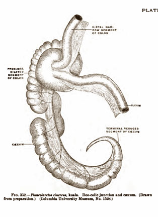
[1788] Book: Marsupials. Edited by Patricia Armati and others. Cambridge University Press, 2006. Chapter 5: "Nutrition and digestion." By Ian D. Hume.
Page 151: "In the koala Phascolarctos cinereus, the closest extant relative to the wombats...."
[1789] Article: "Wombat." Encyclopedia Britannica Ultimate Reference Suite 2004.
"Wombats are ... strictly herbivorous, eating grasses and the inner bark of tree and shrub roots."
[1790] Book: The Anatomy of the Human Peritoneum and Abdominal Cavity: Considered from the Standpoint of Development and Comparative Anatomy. By George S. Huntington. Lea Brothers & Co., 1903. Plate CLXXX. Figure 354:
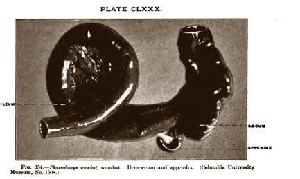
[1791] Paper: "Anatomy of the Koala (Phascolarctos Cincreus)." By A. H. Young. Journal of Anatomy and Physiology: Normal and Pathological, July 1881. Pages 466-474. http://www.pubmedcentral.nih.gov/picrender.fcgi?...
Page 474:
[V]ery short is the caecum in rhizophagous (Wombat) ... it appears that Koala only differs in its visceral anatomy from other Phalangers by the existence of its special gastric glandular apparatus, closely resembling the Wombat (Phascolomys Wombat) in this respect, but differing widely from this animal in the possession of a long caecum, and in the absence of a vermiform appendix.
[1792] Textbook: Introduction to Physical Anthropology. By Lynn Kilgore, Robert Jurmain, Wenda Trevathan. Thomson Wadsworth, 2005.
Page 163: "All gorillas are almost exclusively vegetarian. Mountain gorillas concentrate primarily on leaves, pith and stalk. These foods are also important for western lowland gorillas, but western lowland gorillas also eat considerably more fruit, depending on seasonal availability."
[1793] Article: "Cellulose." Contributor: John Blackwell (Ph.D., Professor of Macromolecular Science, Case Western Reserve University). World Book Encyclopedia, 2007 Deluxe Edition.
"The foods most abundant in cellulose are vegetables that consist of stalks or leaves, such as celery and spinach."
[1794] Paper: "The primate caecum and appendix vermiformis: A comparative study." By G.B.D. Scott. Journal of Anatomy, October 1980. Pages 549-563.
Page 561: "On the other hand, the appendix of the gorilla, as illustrated by Elftman & Atkinson (1950), appears remarkably similar to that of the human, arising as it does abruptly from the posteromedial aspect of the caecum while the three fully formed taeniae coli fuse at its base."
[1795] Book: The Anatomy of the Human Peritoneum and Abdominal Cavity: Considered from the Standpoint of Development and Comparative Anatomy. By George S. Huntington. Lea Brothers & Co., 1903. Plate CCXXXV:
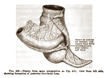
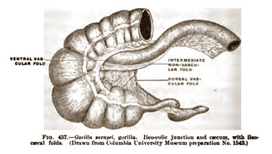
NOTE: Of all the sources I examined, I found no mention of any differentiation between the sizes and shapes of the cecums in the three different types of gorillas, which are the western lowland, the eastern lowland, and the mountain gorilla.
[1796] Article: "Monkey." Encyclopedia Britannica Ultimate Reference Suite 2004.
"Monkeys are arranged into two main groups: Old World and New World."
[1797] Book: The Antecedents of Man: An Introduction to the Evolution of the Primates. By Wilfred E. Le Gros Clark. Quadrangle books, 1971. Page 295:
In the New World monkeys the caecum is conical, non-sacculated and relatively voluminous, tapering gradually to a pointed extremity which may be elongated and thus simulate an appendix (fig. 144A). But it does not contain local accumulations of lymphoid tissue concentrated to the extent which is characteristic of a true vermiform appendix; it is to be regarded, rather, as the pointed end of the caecal pouch proper. In the Old World monkeys the caecum is generally smaller and terminates in a blunt, rounded extremity (fig. 144B). ... The vermiform appendix is completely absent in these monkeys. ...
In the anthropoid apes and man the caecum closely resembles that of the Catarrhine [Old World] monkeys in its relative size and shape.... It differs markedly, however, in having attached to it a narrow, tubular appendix.
NOTE: Figures 144 A-C on page 296 are very instructive. Figure A shows the cecum of a New World monkey, which looks remarkably similar to that of the kangaroo cecum shown on page 226 of Rational Conclusions. From Figures B and C, we see that the cecums of an Old World monkey and a human are almost exactly the same, except for the fact that humans have an appendix.
[1798] Book: The Natural History of the Primates. By J.R. Napier & P.H. Napier. MIT Press, 1985.
Dust cover: "John Napier is Visiting Professor of Primate Biology, Birkbeck College, London. Prior to this appointment he was the first Director of the Primate Biology Program at the Smithsonian Institution. Prue Napier is widely recognized as a leading authority on primate biology."
Page 44:
The vermiform appendix is completely absent in Old World monkeys. In New World monkeys, the lower end of the caecum is conical and tapering, and might be considered to be the homologue of the appendix of the apes and man; however the absence of lymphoid tissue, so characteristic of the human appendix, argues against this hypothesis. The appendix appears to be a specialization of the apes and man, rather than the vestigial organ it is commonly considered.
[1799] Article: "Rabbit." Contributor: Terri McGinnis (D.V.M., Veterinarian). World Book Encyclopedia, 2007 Deluxe Edition.
"In spring and summer, rabbits eat green leafy plants, including clover, grass, and herbs. In winter, they eat the twigs, bark, and fruit of bushes and trees."
[1800] Book: Textbook of Rabbit Medicine. By Frances Harcourt-Brown. Reed Educational and Professional Publishing, 2002.
Page 9: "The thin walled caecum ends in a narrow blind appendix that is heavily endowed with lymphoid tissue."
[1801] Paper: "Rabbit post-mortem technique." By Jill Pearson. Veterinary Times, January 2001. http://www.vetsforum.co.uk/article_post_mortem.htm
NOTE: For a picture of a rabbit cecum and appendix, see the second picture under the heading "The body cavities."
[1802] Abstract: "Digestion and metabolism of a natural foliar diet (Eucalyptus punctata) by an arboreal marsupial, the koala (Phascolarctos cinereus)." By S. J. Cork & others. Journal of Comparative Physiology, June, 1983. http://www.springerlink.com/content/h317p550g1346751/
"The average apparent digestibilities of dietary constituents were ... cellulose 31%...."
[1803] Book: Marsupials. Edited by Patricia Armati and others. Cambridge University Press, 2006. Chapter 5: "Nutrition and digestion." By Ian D. Hume. Pages 151-2:
In the koala Phascolarctos cinereus, the closest extant relative to the wombats, the efficiency of particle size reduction of ingested Eucalyptus leaves depends on the maintenance of sharp cutting edges on the molars.... Aged koalas, with their worn premolars and molars, are unable to comminute eucalyptus leaves finely enough, and eventually die from starvation, often with their stomachs full of coarsely macerated leaves. ... Microbial fermentation is extensive throughout the caecum and proximal colon, but rates of fermentation are low, and the SCFAs [short-chain fatty acids] produced contribute only 9% of the digestible energy intake in captive koalas (Cork et al. 1983). ... Thus by far the most important site of energy absorption in the koala is the small intestine, with the bulk of the energy absorbed coming from cell contents rather than plant cell walls.
[1804] Paper: "Surgical Removal Of The Cecum And Its Effect On Digestion And Growth In Rabbits." Journal of Nutrition, October 1955. Pages 261-270. http://jn.nutrition.org/cgi/reprint/57/2/261.pdf
Page 262: "The cecum was removed, as a rule, when the animal was at least 8 weeks old."
Page 265: "Cecectomized animals always remained outwardly normal in appearance. After extended periods following cecectomy, however, enlargement of the adrenal and colon were observed. The kidneys were enlarged, pale, and slightly scarred. All other organs appeared normal. The enlargement of the colon may have been in compensation for the absence of the cecum."
Page 270: "The digestibility of fiber (cellulose) was equal in both normal and in cecectomized rabbits."
[1805] Book: Histology: A Text and Atlas With Correlated Cell and Molecular Biology. By Michael H. Ross & Wojciech Pawlina. Fifth edition. Lippincott Williams & Wilkins, 2006.
Page 545: "The four layers characteristic of the alimentary canal are present throughout [the large intestine]."
Page 550: "Although the lamina propria of the large intestine contains the same basic components as the rest of the digestive tract, it demonstrates some additional structural features and greater development of some others."
Page 572: "The wall of the appendix is much like that of the small intestine,* having a complete longitudinal layer of muscularis externa, but it lacks both plicae circulares and villi. ... Note that the epithelium of the glands in the appendix is similar to that of the large intestine."
NOTE:
* The appendix is considered part of the large intestine.
[1806] Book: Cell Biology and Histology. By Leslie P. Gartner, James L. Hiatt, Judy M. Strum. Fifth edition. Lippincott Williams & Wilkins, 2006. Page 232:
The epithelium [outside layer] of the mucosa [mucous membranes] of the cecum and colon is simple columnar with numerous goblet cells, surface absorptive cells, and occasional DNES cells. ... The lamina propria [layer just beneath the epithelium] is similar to that of the small intestine, possessing lymphoid nodules, blood and lymph vessels, and closely packed crypts of Lieberkühn, which lack Paneth cells.
Page 233:
Mucosa of the appendix.... The epithelium is simple columnar and contains surface columnar cells and goblet cells. ... The lamina propria displays numerous lymphoid nodules (capped by M cells) and lymphoid cells. It does not form villi but possesses shallow crypts of Lieberkühn with some goblet cells, surface columnar cells, regenerative cells, occasional Paneth cells, and numerous DNES cells, especially deep in the crypts.
NOTE: Among the differences above, note that Paneth cells and M cells are present in the appendix, cells located in the epithelium of the cecum are found in the lamina propria of the appendix, and there are far more DNES cells in appendix. See the following citation for an explanation of Paneth cells.
[1807] Book: Histology: A Text and Atlas With Correlated Cell and Molecular Biology. By Michael H. Ross & Wojciech Pawlina. Fifth edition. Lippincott Williams & Wilkins, 2006.
Page 538: "Paneth Cells, whose primary function is to maintain mucosal innate immunity by secreting antimicrobial substances."
Page 540: "Paneth cells play a role in regulation of normal bacterial flora of the small intestine. ... This antibacterial action and their ability to phagocytose certain bacteria and protozoa suggest that Paneth cells play a role in regulating the normal bacterial flora of the small intestine."
[1808] Paper: "Serrated adenomas of the appendix." By Carlos A. Rubio. Journal of Clinical Pathology, September 2004. Pages 946–949. http://www.pubmedcentral.nih.gov/articlerender.fcgi?...
However, it should be born in mind that there are histofunctional differences between the mucosa of the colon and rectum and that of the appendix. In the appendix, most of the lumen is lined by specialised follicle associated epithelium. That specialised epithelium has—in contrast to the epithelium of the colorectal mucosa—few goblet cells and many of the columnar cells are of the M cell type (that is, membranous cells able to attract luminal antigens). In addition, the mucosa of the appendix contains neuroendocrine complexes (a collection of neuroendocrine ganglia interconnected with neural fibres) just beneath the crypts.
NOTE: In November 2007, I wrote Dr. Rubio to find out of there were "histofunctional differences between the mucosa of the cecum and appendix." After citing the quote above, he explained that the cecum was essentially part of the colon, and that the appendix was histologically and anatomically distinct from both of these organs:
The cecum (latin =blind), being the blind end of the colon, is not at variance with the rest of that organ, except for normally occurring occasional lymphatic aggregates, a phenomenon shared with the rectum. Consequently, the histo-anatomical make-up of the colorectum (including the cecum) is at variance with that of the appendix.
[1809] Book: Anatomy: A Regional Study of Human Structure. By Ernest Gardner, Donald J. Gray, Ronan O'Rahilly. Fourth edition. W.B. Saunders, 1975.
Page 392: "The appendix lacks sacculations and has a longitudinal muscle coat that lacks taeniae."
NOTE: Picture 35-17 on page 391 shows these differences between the appendix and cecum.
[1810] Book: The Human Body: an Introduction to Structure and Function. By Adolf Faller, Michael Schünke, Gabriele Schünke. Thieme, 2004. Translated and revised from the 13th German edition (1999) by Oliver French.
Page 414: "The appendix ... has an important function in the human specific immune system (see above) [page 296]."
Page 296:
Because of their large surface area, the intestines play a central role in immunity. After all, 70-80% of all antibody-producing cells are situated in the intestinal wall. ... Diffuse collections and loose associations of lymphocytes (lymphatic follicles) can be found throughout the gastrointestinal tract, which because of its direct contact with ingested nutrients, is an ideal portal of entry for antigens [toxic substances]. Organized lymphatic tissue is present in the vermiform appendix.... Into the epithelium [lining] of the intestinal mucosa are dispersed specific cells that apparently selectively recognize and take up antigenic substances.
[1811] Textbook: The Science of Evolution. By William D. Stansfield (Professor of Biological Sciences at California Polytechnic State University). Macmillan, 1977. Page 123:
Probably one of the most dramatic lines of evidence for evolution is seen in vestigial or rudimentary structures. These nonfunctional structures and organs are easily explained by the theory of evolution as the now useless remnants of functional structures in ancestral stock. ... Nearly a hundred vestigial structures have been cataloged in modern humans.
NOTE: As examples of vestigial organs, Figure 5.22 shows the nictitating membrane, muscles to move ears, pointed canine [teeth], third molar [wisdom teeth], mammae on male, hair on body, vermiform appendix, pyramidalis muscle, caudal vertebrate, and segmental muscles on the abdomen.
[1812] Textbook: Fundamentals of Anatomy & Physiology. By Frederic H. Martini (Ph.D. in comparative and functional anatomy from Cornell University) Prentice Hall, 2001. Page 857.
[1813] Textbook: Biology. By Kenneth R. Miller & Joseph Levine. Prentice Hall, 1993. Page 284.
Page 284: "Vestigial organs seem to serve no useful purpose at all. ... Structures that are not used may become smaller and smaller but may never disappear completely."
[1814] Textbook: Biology. By Kenneth R. Miller & Joseph Levine. Prentice Hall, 1993. Page 284.
[1815] Book: Finding Darwin's God. By Kenneth R. Miller. Cliff Street Books, 1999. Pages 100-101.
[1816] Book: Controversies in Perinatal Medicine: Studies on the Fetus as a Patient. Edited by José M. Carrera & others. Parthenon Publishing, 2003. Chapter 8: "Early diagnosis of congenital anomalies: 4. Adnexal markers of aneuploidy." By F. Bonilla Musoles & others. Page 91:
The yolk sac has several crucial functions for embryonic development, besides its fundamental role in the metabolism of the embryo. The yolk sac starts hematopoiesis [the formation of blood cells], neoangiogenesis [the formation of blood vessels] and the first blood circulation of the embryo. It is responsible for maternal-embryo gaseous, electrolyte, ion and hormonal exchanges in addition to all other exchanges necessary for embryo development. It contributes to the development of the midgut [middle portion of digestive tract] and to abdominal wall closure. It also has an important role in fetal immunology by producing certain transitory proteins....
Page 92: "It is indisputable that the yolk sac has a critical role in organogenesis [the formation of organs] and normal embryo development."
[1817] Textbook of Fetal Ultrasound. Edited by Richard Jaffe & The-Hung Bui. Parthenon Publishing, 1999. Chapter 4: "Fetal biometry and gestational age estimation." By Bobbi Stebbins & Richard Jaffe.
Page 47: "Normal yolk sac size and shape are associated with good pregnancy outcome whereas abnormal yolk sac size and shape are often a first sign of abnormal embryonic development. Absence of a yolk sac in the presence of a visible embryo is abnormal and associated with subsequent embryonic death."
[1818] Book: An Introduction to Human Disease: Pathology and Pathophysiology Correlations. By Leonard V. Crowley. Seventh edition. Jones and Bartlett, 2007.
Page 466: "Part of the yolk also becomes enfolded within the embryo when flexion occurs (Figure 18-5). The enclosed part will give rise to the intestinal tract and other important structures."
[1819] Book: Color Atlas of Life Before Birth. By Marjorie A. England. Year Book Medical Publishers, 1983. Page 44:
As the extra-embryonic coelom forms the primary yolk sac degenerates and a smaller secondary yolk sac forms lined by endoderm. The first vascular blood forms from yolk sac mesoderm (Horizon VI, Day 13). This will provide embryonic blood until the liver starts to form blood (Week 5). It also provides nutrients during Week 2 and 3 while the chorioallantoic placenta develops. During Week 3 the primordial germ cells formed on the yolk sac migrate into the embryo. In Week 4 the body folds constrict the yolk sac and the portion incorporated into the embryo forms the epithelium of the gut tube.
[1820] Book: An Atlas of the Blood and Bone Marrow. By R. Philip Custer (Emeritus Professor of Pathology, The University of Pennsylvania School of Medicine; Senior Member, The Institute for Cancer Research, Fox Chase; Consultant, The Presbyterian-University of Pennsylvania Medical Center). Second edition. W. B. Saunders Company, 1974. Page 9:
All formed elements of the blood take their origin from embryonal connective tissue, the mesenchyme, generally through primitive blood cells. In the primitive-streak and somite stages of the embryo, these cells are found exclusively in the yolk sac during the formation of blood islands in the area vasculosa, coincident with the development of the earliest blood vessels.
[1821] Book: Anatomy and Physiology for Midwives. By Jane Coad with Melvyn Dunstall. Mosby, 2001.
Page 172: "The allantois and yolk sac are semivestigial structures that have a more important role in other species, such as birds and reptiles, where the yolk sac is important in nutrition of the maternally isolated eggs and the allantois has a respiratory and excretory role."
[1822] Article: Was Darwin Wrong? By David Quammen. National Geographic, November 2004 (cover story). http://ngm.nationalgeographic.com/ngm/0411/feature1/fulltext.html
Vestigial characteristics are still another form of morphological evidence [for evolution], illuminating to contemplate because they show that the living world is full of small, tolerable imperfections. Why do male mammals (including human males) have nipples? ... Darwin raised all these questions, and answered them, in The Origin of Species. Vestigial structures stand as remnants of the evolutionary history of a lineage.
[1823] Book: The Impacted Lower Wisdom Tooth. By A. J. MacGregor. Oxford University Press, 1985. Page 3:
At it most simple the idea is that as the successive teeth erupt the jaws grow to make room for them. If the jaws are not big enough then there will not be room for all of the teeth, and the last to erupt will become misplaced. This hypothesis is quite satisfactory because it not only explains why third molars, both upper and lower become impacted, it also explains why deciduous teeth do not.
Pages 3-16 analyze evidence pertaining to why this occurs and conclude with these words:
Evidence derived from palaeontology, anthropology, and experiment indicates very convincingly that a reduction in jaw size has occurred due to civilization. The main associated factor appears to be the virtual absence of interproximal attrition, but initial tooth size may have had some effect. Jaw size and dental attrition are related and they have both decreased with modern diet. Jaws were thought to be reduced in size in the course of evolution but close examination reveals that within the species Homo sapiens, this may not have occurred. What was thought to be a good example of evolution in progress has been shown to be better explained otherwise.
[1824] Abstract: "The Prophylactic Extraction of Third Molars: A Public Health Hazard." By Jay W. Friedman. American Journal of Public Health, September 2007. Pages 1554-1559. http://www.ajph.org/cgi/content/abstract/97/9/1554
Ten million third molars (wisdom teeth) are extracted from approximately 5 million people in the United States each year at an annual cost of over $3 billion.
In addition, more than 11 million patient days of "standard discomfort or disability"—pain, swelling, bruising, and malaise—result postoperatively, and more than 11000 people suffer permanent paresthesia—numbness of the lip, tongue, and cheek—as a consequence of nerve injury during the surgery. At least two thirds of these extractions, associated costs, and injuries are unnecessary, constituting a silent epidemic of iatrogenic injury that afflicts tens of thousands of people with lifelong discomfort and disability.
[1825] Book: The Human Body: an Introduction to Structure and Function. By Adolf Faller, Michael Schünke, Gabriele Schünke. Thieme Medical Publishers, 2004. Translated and revised from the 13th German edition (1999) by Oliver French, M.D. Page 672.
[1826] Textbook: Fundamentals of Anatomy & Physiology. By Frederic H. Martini (Ph.D. in comparative and functional anatomy from Cornell University) Prentice Hall, 2001. Pages 152-3.
[1827] Book: Atlas of the Sensory Organs: Functional and Clinical Anatomy. Edited by András Csillag. Humana Press, 2005. Chapter 5: "The Skin and Other Diffuse Sensory Systems." By Andrea D. Székely & András Csillag. Page 213.
[1828] Book: di Fiore's Atlas of Histology with Functional Correlations. By Victor P. Eroschenko. Tenth Edition. Lippincott, Williams & Wilkins, 2005. Page 198.
[1829] Textbook: Fundamentals of Anatomy & Physiology. By Frederic H. Martini (Ph.D. in comparative and functional anatomy from Cornell University) Prentice Hall, 2001. Page 219.
[1830] Book: Anatomy of the Human Body (Gray's Anatomy). By Henry Gray. Edited by Charles Mayo Goss. 29th American edition. Lea & Febiger, 1973. Page 118.
[1831] Paper: "A Comparative Study of the Nictitating Membrane of Birds and Mammals." By E. Philip Stibbe. Journal of Anatomy, 1928. Pages 159–176. http://www.pubmedcentral.nih.gov/picrender.fcgi?artid=1250027&blobtype=pdf